
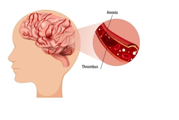
A brain hematoma is an accumulation of blood inside the skull. It may occur within the brain tissue itself or between the membranes surrounding the brain as a result of bleeding from a blood vessel.
Yes. A brain hematoma may cause persistent or recurrent headaches, particularly in some cases of chronic subdural hematoma.
Yes. A brain hematoma may cause dizziness and balance problems. These symptoms depend on the location and size of the hematoma and the extent to which it affects the parts of the brain responsible for controlling balance and movement.
Yes. A brain hematoma may affect memory, concentration, and attention, depending on its location, size, and impact on brain function. These abilities may gradually improve after treatment, and patients may benefit from neurological rehabilitation programs.
Yes. A brain hematoma can be fatal in severe cases, particularly if it causes significant bleeding, dangerous pressure on brain tissue, or a severe increase in intracranial pressure.
Yes. In some cases, a brain hematoma may lead to a coma, especially when the bleeding is severe or causes increased intracranial pressure, affecting brain function and the level of consciousness.
Yes. Bleeding into the brain's ventricles may cause hydrocephalus if it obstructs the flow or absorption of cerebrospinal fluid (CSF), resulting in fluid accumulation within the brain's cavities.
Head Injuries: Head injuries are among the most common causes of intracranial hematomas. They may result from falls, car accidents, blows to the head, or severe impacts.
High Blood Pressure: Severely elevated or uncontrolled blood pressure may cause a blood vessel in the brain to rupture, resulting in bleeding and hematoma formation.
Brain Aneurysm: This occurs when a weakened area of an artery wall in the brain bulges outward. The aneurysm may rupture and cause bleeding inside the skull.
Vascular Malformations: These include arteriovenous malformations (AVMs), which involve abnormal connections between arteries and veins and may lead to bleeding in the brain.
Blood Clotting Disorders: Conditions such as a low platelet count or abnormalities in clotting factors can reduce the body's ability to stop bleeding, increasing the risk of hematoma formation.
Use of Anticoagulant Medications: Certain blood-thinning medications, such as warfarin and other anticoagulants, may increase the risk of bleeding inside the skull, particularly when doses are not properly managed or other risk factors are present.
Brain Tumors: Some brain tumors may bleed within the tumor tissue or in the surrounding areas, resulting in the formation of a hematoma.
Age-Related Changes: As people age, certain blood vessels and veins surrounding the brain may become more vulnerable to tearing, increasing the risk of hematoma formation, particularly subdural hematomas.
Use of Stimulant Drugs: Substances such as cocaine and amphetamines may cause a sudden increase in blood pressure, raising the risk of blood vessel rupture and bleeding.
Severe Liver Disease: Certain liver conditions may interfere with the production of blood-clotting factors, making the body more susceptible to bleeding.
Epidural Hematoma: This usually occurs following a head injury that causes one of the arteries or blood vessels located between the skull and the dura mater to rupture.
Subdural Hematoma: This is usually caused by the rupture of small veins that connect the surface of the brain to the surrounding membranes. It may occur after a minor head injury, particularly in older adults.
Intracerebral Hemorrhage: This occurs when a blood vessel ruptures within the brain tissue. It may be associated with high blood pressure, vascular malformations, or blood clotting disorders.
Subarachnoid Hemorrhage: This occurs when blood leaks into the space between the arachnoid mater and the pia mater. It may result from a ruptured brain aneurysm or a head injury.
1. Epidural Hematoma
An epidural hematoma is an accumulation of blood between the skull bones and the outer membrane surrounding the brain, known as the dura mater. It usually occurs following a head injury that causes a blood vessel to rupture and may lead to increased intracranial pressure.
2. Subdural Hematoma
A subdural hematoma is an accumulation of blood in the space between the dura mater and the arachnoid membrane surrounding the brain. It is usually caused by the rupture of veins that connect the brain's surface to the surrounding membranes. It may occur following a head injury, even a minor one, particularly in older adults.
3. Subarachnoid Hemorrhage
A subarachnoid hemorrhage is bleeding into the space between the arachnoid mater and the pia mater, two of the membranes surrounding the brain. It may result from a ruptured brain aneurysm or a head injury and is often characterized by the sudden onset of a severe headache.
4. Intracerebral Hematoma
An intracerebral hematoma is an accumulation of blood within the brain tissue itself due to the rupture of a blood vessel. It may be associated with high blood pressure, vascular malformations, or blood clotting disorders.
5. Intraventricular Hemorrhage
An intraventricular hemorrhage is bleeding inside the brain's ventricles, which are cavities within the brain that contain cerebrospinal fluid (CSF). Blood accumulation in these cavities may obstruct the flow of CSF and potentially lead to hydrocephalus.
Subdural hematomas can be classified into three main types according to the time elapsed since the bleeding occurred:
| Type | Approximate Duration |
|---|---|
| Acute Subdural Hematoma | Within the first 72 hours |
| Subacute Subdural Hematoma | From 3 days to 3 weeks |
| Chronic Subdural Hematoma | More than 3 weeks after onset |
The main symptoms and signs that may accompany an intracranial hematoma include:
Severe or Persistent Headache: The headache may gradually become more intense or appear suddenly, depending on the nature of the bleeding.
Dizziness and Balance Problems: The patient may have difficulty standing or walking normally.
Nausea and Vomiting: Vomiting may be recurrent, particularly when intracranial pressure increases.
Vision Problems: These may include blurred vision, double vision, or loss of part of the visual field.
Muscle Weakness or Numbness: These symptoms may affect an arm or leg or involve one side of the body.
Difficulty Walking and Moving: This may occur when the areas of the brain responsible for controlling movement and balance are affected.
Speech Difficulties: These may include difficulty speaking, expressing thoughts, or understanding speech.
Excessive Drowsiness and Lethargy: The patient may feel unusually sleepy and have difficulty staying alert.
Memory and Concentration Problems: The patient may have difficulty remembering information or maintaining attention.
Behavioral and Personality Changes: These may include irritability, confusion, or changes in usual behavior.
Loss of Consciousness or Fainting: This may occur in cases where brain function is significantly affected.
Seizures: These may occur as a result of disruption to the brain's normal electrical activity.
The specialist, whether a neurologist or a neurosurgeon, begins by assessing the patient's overall health and performing a comprehensive neurological examination, which includes:
Assessment of Consciousness and Alertness: To determine the patient's ability to respond to stimuli, understand questions, and follow instructions.
Muscle Strength and Movement Examination: By assessing arm and leg movements and checking for weakness or paralysis.
Sensory Examination: To identify any numbness or loss of sensation in specific parts of the body.
Pupil Examination: To check the size of the pupils and how they respond to light.
Assessment of Speech and Comprehension: To identify difficulties with speaking, expressing thoughts, or understanding speech.
Balance and Coordination Assessment: To evaluate the patient's ability to move and walk normally.
Detection of Seizures and Neurological Signs: To identify any neurological disturbances associated with the hematoma.
Imaging studies are among the most important tools for diagnosing intracranial hematomas and determining the location and size of the bleeding, as well as its impact on the brain.
The primary imaging test in acute and emergency cases
A non-contrast CT scan is usually one of the first tests performed when intracranial bleeding is suspected. It helps to:
Detect the presence of bleeding or a hematoma.
Determine the location and size of the hematoma.
Assess the degree of pressure on brain tissue.
Detect brain swelling or midline shift.
Identify signs of hydrocephalus.
MRI provides detailed images of brain tissue and may help to:
Detect hematomas and determine their characteristics.
Evaluate the stages of bleeding and estimate its age in certain cases.
Identify some vascular abnormalities and tumors that may be responsible for the bleeding.
Investigate cases in which CT scan findings are inconclusive.
Evaluate certain cases of chronic hematoma.
This test is used to visualize the blood vessels in the brain after injecting a contrast agent. It helps to:
Detect brain aneurysms.
Identify certain arteriovenous malformations.
Determine the possible source of bleeding.
Evaluate abnormal changes in the brain's arteries.
This test helps evaluate the blood vessels inside the brain and investigate possible vascular causes of bleeding, such as aneurysms and vascular malformations.
Diagnostic cerebral angiography is a highly detailed imaging procedure used to examine the blood vessels in the brain. Doctors may request it in specific cases, particularly when an aneurysm or arteriovenous malformation is suspected, or when previous imaging tests require further clarification.
The doctor may request several laboratory tests to assess the patient's overall condition and identify any disorders that could increase the risk of bleeding or affect treatment decisions.
| Laboratory Test | Purpose |
|---|---|
| Complete Blood Count (CBC) | Measures hemoglobin levels and platelet counts and evaluates blood components. |
| Coagulation Tests (PT/INR and aPTT) | Assess the blood's ability to clot and identify clotting disorders. |
| Liver Function Tests | Detect liver disorders that may affect the production of clotting factors. |
| Kidney Function Tests | Evaluate kidney function, particularly before certain imaging tests and treatments. |
| Blood Glucose Test | Detect blood sugar abnormalities and assess the patient's overall condition. |
The doctor determines the appropriate medication based on the patient's condition and the cause of bleeding. The main medication groups used include:
| Medication Type | Purpose |
|---|---|
| Blood Pressure-Lowering Medications | Control high blood pressure and reduce the risk of continued or worsening bleeding. |
| Anticoagulant Reversal Agents | Reverse the effects of certain blood-thinning medications when bleeding is associated with their use. |
| Anticonvulsant Medications | Treat seizures or prevent them in selected cases when medically indicated. |
| Medications to Reduce Intracranial Pressure | Manage increased pressure inside the skull when urgent intervention is required. |
| Pain Relievers | Relieve headaches and pain according to the doctor's assessment. |
| Antiemetic Medications | Control nausea and vomiting associated with the condition. |
These medications are used to control high blood pressure, particularly in cases of bleeding associated with hypertension. Examples include:
Labetalol.
Nicardipine.
The doctor determines the appropriate medication and the rate at which blood pressure should be lowered based on the type of bleeding and the patient's condition, while avoiding an excessive or rapid reduction in blood pressure.
Patients who develop intracranial bleeding while taking blood-thinning medications may need specific reversal agents to counteract their effects and reduce the risk of continued bleeding. Examples include:
Vitamin K with Prothrombin Complex Concentrate (PCC): Used to reverse the effects of warfarin in cases of warfarin-associated bleeding.
Idarucizumab: A medication specifically used to reverse the effects of dabigatran.
Andexanet Alfa: May be used to reverse the effects of certain factor Xa inhibitors, depending on medical assessment and availability.
Protamine: Used to reverse the effects of heparin in specific cases.
The choice of medication depends on the type of anticoagulant the patient is taking, the time of the last dose, laboratory test results, and the severity of the bleeding.
The doctor may prescribe anticonvulsant medications if the patient experiences seizures or if medical findings indicate that their use is necessary. Examples include:
Levetiracetam.
Fosphenytoin.
In some cases, a hematoma may cause increased intracranial pressure, requiring specific medications to lower the pressure and protect brain tissue. Examples include:
Mannitol.
Hypertonic Saline.
These treatments are used in selected cases under close monitoring in the hospital, with regular assessment of blood pressure, kidney function, and blood electrolyte levels.
It is important to understand that these medications do not dissolve the hematoma itself. Instead, they help manage some of the complications caused by it.
The choice of surgical procedure depends on several factors, including:
The location of the intracranial hematoma.
The size and extent of the hematoma.
The type of bleeding and how long ago it occurred.
The degree of pressure exerted on brain tissue.
The patient's level of consciousness and neurological condition.
The underlying cause of the bleeding and whether it is ongoing or has stopped.
How Is the Procedure Performed?
The patient is placed under general anesthesia.
A surgical incision is made in the scalp.
A section of the skull bone is opened to access the affected area.
The location of the hematoma is identified, and the accumulated blood is removed.
The source of bleeding is addressed and controlled whenever possible.
The bone flap is repositioned and secured, and the surgical wound is closed.
When Is It Used?
This procedure is used in selected cases of acute epidural and subdural hematomas, as well as certain cases of intracerebral hemorrhage, particularly when the hematoma is large or causes significant pressure on the brain.
This is one of the common procedures used to treat chronic subdural hematomas. It aims to drain the accumulated blood through small openings made in the skull.
How Is the Procedure Performed?
The patient receives anesthesia, either local or general, depending on their condition.
One or more small holes are drilled into the skull.
A thin drainage tube is inserted to remove the accumulated blood.
The cavity may be irrigated when necessary, according to the surgeon's assessment.
A temporary drainage tube may be left in place in some cases and removed once the patient's condition is stable.
When Is It Used?
It is commonly used for chronic subdural hematomas, particularly when they cause neurological symptoms or exert pressure on brain tissue.
This procedure is performed in certain severe cases involving dangerously increased intracranial pressure that does not respond to appropriate medical treatment.
How Is the Procedure Performed?
A surgical incision is made in the scalp.
A section of the skull bone is removed to create additional space for the brain.
The membrane surrounding the brain may be expanded when necessary through a procedure known as duraplasty.
This allows swollen brain tissue to expand without being subjected to further dangerous pressure.
The scalp is closed, and the removed bone flap is preserved for possible reattachment at a later stage.
When Is It Used?
It is used in certain critical cases involving severe brain swelling or persistently elevated intracranial pressure. It may be performed alongside hematoma evacuation when clinically necessary.
Endoscopic evacuation is a minimally invasive surgical technique used in selected cases to remove accumulated blood through a relatively small opening in the skull.
How Is the Procedure Performed?
A small opening is made in the skull.
A surgical endoscope equipped with a small camera is inserted.
The endoscope is guided toward the location of the hematoma.
The accumulated blood is suctioned out and removed using specialized surgical instruments.
The affected area is examined, and efforts are made to control the bleeding.
When Is It Used?
It may be used in selected cases of intracerebral hemorrhage, depending on the location and size of the hematoma, the suitability of the technique, and the surgical team's experience.
This is a minimally invasive procedure used in certain cases of intracerebral hematoma. It relies on imaging guidance to determine a precise route to the site of the bleeding.
How Is the Procedure Performed?
The location of the hematoma is identified using a CT scan or other appropriate imaging techniques.
A precise trajectory to the hematoma is calculated.
A specialized surgical needle or catheter is inserted through a small opening in the skull.
Part of the accumulated blood is aspirated.
A drainage tube may be inserted when necessary. In selected cases, clot-dissolving medications may be administered into the hematoma according to specialized medical protocols.
When Is It Used?
It may be considered in certain cases of intracerebral hemorrhage. The decision depends on the location and size of the bleeding, the nature of the patient's condition, and the potential risks.
This is an interventional technique used to treat certain cases of chronic subdural hematoma. It aims to reduce blood flow to the membranes and blood vessels associated with the hematoma, helping limit its persistence or recurrence.
How Is the Procedure Performed?
A thin catheter is inserted through an artery, usually in the groin or wrist.
Using imaging guidance, the catheter is advanced until it reaches the middle meningeal artery.
A specialized embolic material or another suitable method is used to block the targeted branches of the artery.
The completion of the procedure is confirmed through angiographic imaging.
When Is It Used?
It is used in selected cases of chronic subdural hematoma. It may be performed alongside surgical drainage or as a standalone treatment in certain patients, depending on the specialist's assessment.
This procedure is used to drain cerebrospinal fluid and blood from the brain's ventricles when necessary, particularly in cases of intraventricular hemorrhage accompanied by hydrocephalus or increased intracranial pressure.
How Is the Procedure Performed?
A small opening is made in the skull.
A thin catheter is inserted into one of the brain's ventricles.
The catheter is connected to an external drainage system.
Blood and cerebrospinal fluid are drained at a rate determined by the medical team.
Intracranial pressure and the volume of drained fluid are monitored.
When Is It Used?
It is used in certain cases of intraventricular hemorrhage, particularly when the bleeding obstructs the flow of cerebrospinal fluid and causes hydrocephalus.
This procedure is used to treat certain cases of brain aneurysms, particularly when aneurysm rupture is responsible for the bleeding.
How Is the Procedure Performed?
The patient is placed under general anesthesia.
The skull is opened to access the affected artery.
The neck of the aneurysm is identified.
A small metal clip is placed across the aneurysm's neck to prevent blood from flowing into it.
The surgeon ensures that normal blood flow through the artery and nearby blood vessels is maintained.
When Is It Used?
It is used when bleeding results from a ruptured aneurysm. The choice of this procedure depends on the aneurysm's location and shape, as well as the patient's condition.
This is an endovascular technique used to treat brain aneurysms without the need for open skull surgery. It aims to isolate the aneurysm from the bloodstream.
How Is the Procedure Performed?
A thin catheter is inserted through an artery, usually in the groin or wrist.
The catheter is guided through the blood vessels until it reaches the arteries in the brain.
A microcatheter is advanced into the aneurysm.
Small platinum coils are placed inside the aneurysm to reduce blood flow into it and isolate it from the circulation.
The outcome is confirmed using angiographic imaging.
When Is It Used?
It is used in selected cases of brain aneurysms and may serve as an alternative to open surgery, depending on the aneurysm's location and shape, the patient's condition, and specialist assessment.
This procedure is used in certain cases of hematomas located in the posterior fossa of the skull, particularly when the bleeding puts pressure on the cerebellum or brainstem.
How Is the Procedure Performed?
A surgical incision is made in the back of the head.
A section of the posterior skull bone is removed to access the affected area.
The location of the hematoma is identified.
The accumulated blood is removed, reducing pressure on the cerebellum and brainstem.
Any associated complications are managed according to the patient's condition.
When Is It Used?
It is used in selected cases of hemorrhage and hematomas located in the posterior part of the brain, particularly when they compress the cerebellum or brainstem or cause hydrocephalus.
This surgical procedure is used to treat certain cases of brain arteriovenous malformations (AVMs). An AVM is an abnormal connection between arteries and veins that may lead to bleeding.
How Is the Procedure Performed?
The location of the vascular malformation is identified using specialized imaging tests.
The surgeon accesses the affected area.
The abnormal blood vessels are identified and disconnected from the circulation.
The vascular malformation is removed while preserving healthy blood vessels and surrounding brain tissue as much as possible.
The surgeon confirms that the malformation has been removed through surgical assessment and imaging.