

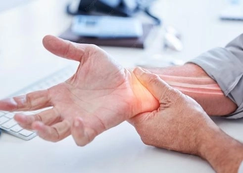
If you experience wrist pain after a fall or injury—even if it seems minor at first—you may be dealing with a more significant condition than expected: a scaphoid fracture.A scaphoid fracture is one of the most common wrist fractures, yet it requires special attention because it can be difficult to diagnose in the early stages. In many cases, the fracture may not be clearly visible on standard X-rays. If left untreated or if treatment is delayed, it can lead to complications such as delayed bone healing, nonunion, or permanent limitations in wrist movement.In this article by Dalili Medical, we explain what a scaphoid fracture is, its most common causes and symptoms, the potential complications of neglecting treatment, and the available treatment options, including casting, medications, and surgery.Our goal is to help you better understand this injury and ensure that it is managed properly from the very beginning.
A scaphoid fracture is a crack or break in a small bone located in the wrist known as the scaphoid bone. This bone is one of the most important bones in the wrist, as it plays a key role in connecting the bones of the hand to those of the forearm. It also contributes significantly to wrist and thumb movement and helps maintain hand stability.
Located on the thumb side of the wrist.
Part of the group of small carpal bones in the wrist.
Despite its small size, it plays a crucial role in maintaining normal hand movement and stability.
A scaphoid fracture is not always serious; however, it can become problematic if not treated promptly. Potential complications include:
Failure of the fracture to heal properly.
Permanent weakness or reduced function of the wrist.
Early development of wrist arthritis.
For this reason, early diagnosis and treatment are extremely important.
The duration of immobilization depends on the type and location of the fracture:
Simple fractures: Usually require a cast for 6–8 weeks.
Displaced fractures or fractures involving the proximal pole: May require immobilization for 10–12 weeks or longer.
Yes, surgery may be necessary in certain cases when cast treatment alone is insufficient, such as:
Displaced fractures.
Unstable fractures.
Failure of the bone to heal with conservative treatment.
In these situations, surgery is performed to realign and stabilize the bone to promote proper healing.
Yes, some parts of the arm and hand can still be moved, but within certain limits:
The fingers, elbow, and shoulder can usually be moved normally.
The injured wrist should not be moved until the fracture has healed properly.
The following symptoms may indicate a problem:
Persistent pain or pain that worsens over time.
Significant swelling around the injured area.
Numbness or tingling in the fingers.
Lack of noticeable improvement during the treatment period.
In most cases, the impact is temporary, and patients need time off or modified activities during treatment. With appropriate management, most people are able to return to their normal daily activities and work without long-term limitations.
Yes. This condition is known as nonunion, and it is more likely to occur in the following situations:
Delayed diagnosis of the injury.
Inadequate immobilization or stabilization of the fracture.
Smoking, which reduces the body's ability to heal bone effectively.
The scaphoid bone can fracture in different regions:
Occurs near the forearm.
Considered the most serious type due to its limited blood supply.
Has a higher risk of delayed union, nonunion, and avascular necrosis.
Occurs in the middle portion of the scaphoid.
The most common type, accounting for more than 70% of cases.
Most often results from a fall onto an outstretched hand.
Occurs near the thumb side of the scaphoid.
Generally has a better blood supply and tends to heal more easily than other types.
The bone is fractured but remains in its normal anatomical position.
Most cases can be treated successfully with cast immobilization.
The fractured bone fragments have shifted from their normal position.
Surgical fixation is often required to restore proper alignment and stability.
The bone fragments remain in their normal position.
Healing rates are generally better, with a lower risk of complications.
Movement exists between the fracture fragments.
Requires more precise treatment and stronger fixation to achieve successful healing.
A very small crack in the bone.
May not be visible on standard X-rays during the early stages.
Delayed or failed healing of the fracture.
Occurs when the broken bone does not unite despite an adequate healing period.
Bone tissue death caused by an inadequate blood supply.
Most commonly affects the proximal pole of the scaphoid due to its limited vascularity.
A scaphoid fracture is one of the most common wrist fractures and usually occurs as a result of a sudden force or direct pressure applied to the hand, particularly during a fall.
This is the most common cause.
When a person falls, they instinctively extend their hand to break the fall. The wrist is forced into hyperextension, placing significant stress on the scaphoid bone, which may result in a fracture.
Certain sports increase the risk of a scaphoid fracture, including:
Football
Gymnastics
Skiing
Basketball
These activities involve frequent falls or direct impacts that can injure the wrist.
Scaphoid fractures may occur during car, motorcycle, or bicycle accidents, especially when a person instinctively extends their hand during impact.
Examples include:
A strong blow to the wrist.
A heavy object falling onto the hand.
Such injuries can cause a direct fracture of the scaphoid bone.
Although less common, certain conditions increase susceptibility to fractures, including:
Calcium deficiency.
Vitamin D deficiency.
Osteoporosis, particularly in older adults.
Repeated and continuous wrist movements associated with certain occupations or sports may gradually produce a stress fracture within the scaphoid bone over time.
Accurate diagnosis of a scaphoid fracture is extremely important because this injury may not be clearly visible during its early stages. Delayed diagnosis can result in delayed treatment and long-term complications.
The physician will obtain detailed information about the injury, including:
Was there a fall onto an outstretched hand?
What was the mechanism of injury (sports injury, accident, or direct blow)?
When did the pain begin, where is it located, and how severe is it?
The most important historical clue is a fall onto an outstretched hand.
One of the most important clinical signs of a scaphoid fracture.
Gentle pressure over this area produces significant tenderness.
Particularly over the location of the scaphoid bone.
Pain typically worsens with wrist use and hand activities.
Some patients experience weakness when gripping objects.
Usually the first imaging study performed.
However, the fracture may not be visible during the first few days after injury.
For this reason, the physician may request:
X-rays from multiple wrist angles.
Repeat X-rays if symptoms persist despite normal initial imaging.
CT scans provide highly detailed images of the fracture and help determine:
Whether displacement is present.
The exact configuration and extent of the fracture.
MRI is considered the most accurate tool for early diagnosis.
It can:
Detect fractures even when plain X-rays appear normal.
Evaluate the blood supply to the scaphoid bone, which is important in assessing the severity and prognosis of the injury.
May be used when MRI is unavailable.
Can detect increased bone activity and early healing changes in the affected area.
Complications are one of the main reasons why scaphoid fractures require prompt diagnosis and treatment. The scaphoid has a unique blood supply, and delays in treatment can lead to long-term wrist dysfunction.
Nonunion refers to failure of the fracture to heal after the expected healing period.
It most commonly occurs when:
Diagnosis is delayed.
The fracture is not adequately immobilized or stabilized.
Chronic wrist pain.
Reduced wrist and hand function.
Progressive loss of wrist mobility.
Delayed union occurs when the fracture eventually heals but takes longer than the expected healing period.
In such cases, the patient may require:
A longer period of cast immobilization.
Surgical intervention in selected cases.
This is considered one of the most serious complications of a scaphoid fracture.
It occurs when the blood supply to the fractured segment becomes insufficient, particularly in fractures involving the proximal pole of the scaphoid.
Death of a portion of the bone tissue.
Significant impairment of wrist function.
Persistent pain and difficulty with wrist movement.
Wrist arthritis may develop as a result of abnormal joint mechanics caused by the fracture or improper fracture healing.
Chronic wrist pain.
Stiffness and reduced range of motion.
Difficulty performing daily activities.
Patients may experience:
Difficulty holding or grasping objects.
Reduced hand strength.
Limited wrist movement.
These problems may result from joint stiffness or changes in bone alignment following the injury.
Carpal collapse is typically seen in neglected or inadequately treated cases.
Alteration of the normal wrist anatomy.
Partial loss of joint function.
Progressive deterioration of wrist movement over time.
Medications alone are not sufficient to treat a scaphoid fracture. However, they play an important supportive role in relieving pain, reducing inflammation, and promoting recovery alongside immobilization or surgical treatment.
Medications are considered supportive therapy only.
The primary treatment consists of:
Cast immobilization.
Surgical fixation when indicated.
Often considered the first-line option for pain control.
Generally safe for most patients when used as directed.
Effective for mild to moderate pain.
Provides pain relief with minimal gastrointestinal irritation compared with many anti-inflammatory medications.
Examples include:
Ibuprofen.
Diclofenac.
Naproxen.
Reduce pain.
Help control swelling and inflammation around the wrist.
Should generally be taken with food.
May not be suitable for individuals with stomach ulcers or certain kidney conditions.
Long-term use should be supervised by a healthcare professional.
In selected cases, physicians may recommend:
Anti-inflammatory medications.
Supportive treatments such as cold compresses and topical therapies.
An essential mineral for bone formation and strength.
Enhances the body's ability to absorb calcium.
May be recommended in certain cases to support bone metabolism and healing.
Although these supplements do not directly heal the fracture, they may help optimize recovery, particularly in patients with nutritional deficiencies.
Short-term use of stronger analgesics may be prescribed when necessary.
Preventive antibiotics may be used based on the surgeon's clinical judgment.
Medications alone cannot heal a scaphoid fracture without proper stabilization.
Excessive use of pain medications may increase the risk of stomach or kidney complications.
The wrist must remain immobilized, and unnecessary movement should be avoided during the healing period.
Treatment of a scaphoid fracture depends on the location, severity, and stability of the fracture. Management is generally based on three main pillars: medications, cast immobilization, and surgical intervention when required.
Medications do not directly repair the fracture, but they help relieve symptoms and support recovery.
Commonly used medications include:
Paracetamol (Acetaminophen): For pain relief.
Nonsteroidal Anti-Inflammatory Drugs (NSAIDs) such as ibuprofen and diclofenac: To reduce pain and swelling.
Calcium and Vitamin D supplements: To support bone healing.
The cornerstone of treatment for a scaphoid fracture is proper immobilization or stabilization of the bone, not medication alone.
Non-surgical treatment is typically recommended for:
Non-displaced fractures.
Stable fractures.
Application of a thumb spica cast, which immobilizes the wrist and thumb.
Immobilization is generally maintained for 6 to 12 weeks, depending on the fracture location and severity.
Regular follow-up imaging is performed to monitor fracture healing.
Surgery may be recommended in the following situations:
Displaced fractures.
Unstable fractures.
Failure of fracture healing with cast treatment.
Delayed diagnosis.
Development of avascular necrosis.
The fractured bone is stabilized using a screw inserted through a minimally invasive approach without the need for a large surgical incision.
A very small incision is made.
A specialized screw is inserted across the fracture site to stabilize the bone fragments.
Intraoperative imaging is used to guide accurate screw placement.
Minimally invasive procedure.
Less postoperative pain.
Faster recovery and return to activities.
A small surgical incision is made to directly visualize the fracture, restore normal bone alignment, and secure the fragments with internal fixation.
A small incision is created over the wrist.
The fracture fragments are realigned (reduction).
The bone is stabilized using a specialized screw, such as a Herbert screw or a headless compression screw.
Precise restoration of bone alignment.
Particularly suitable for displaced or unstable fractures.
Bone grafting may be necessary in cases of:
Long-standing fractures.
Nonunion.
A gap between fracture fragments.
Bone is harvested from another area of the body.
The graft is placed at the fracture site to stimulate healing.
Bone is transferred together with its blood supply.
Commonly used for avascular necrosis or fractures with poor vascularity.
Generally associated with higher success rates in complex cases.
Removal of the proximal row of wrist bones.
Intended to reduce pain and improve overall wrist function.
Permanent fusion of wrist bones.
Significantly reduces pain but eliminates or severely limits wrist motion.
Reserved for severe or advanced cases.
Following surgery, patients typically require:
Temporary cast or splint immobilization.
Gradual initiation of physical therapy.
Follow-up imaging to confirm bone healing.
Avoidance of heavy lifting until recovery is complete.
Exercise should not begin during the early phase of treatment while the fracture is still healing or while a cast is in place. Premature movement may delay healing or compromise treatment outcomes.
Exercises should only be started after cast removal or with the approval of the treating physician.
Even while the wrist is immobilized, it is important to maintain mobility in the unaffected joints.
Slowly open and close the fingers.
Repeat 10–15 times.
Perform several times each day.
Gently make a fist and then relax the hand.
Avoid excessive force.
Helps prevent stiffness of muscles and tendons.
Bend and straighten the elbow.
Perform gentle circular shoulder movements.
Helps prevent stiffness throughout the upper limb.
Once the cast has been removed, wrist mobility can be gradually restored.
Slowly move the wrist upward and downward.
Begin with a limited and comfortable range of motion.
Gently move the wrist from side to side.
Avoid movements that cause significant pain.
Perform slow circular movements of the wrist.
Helps improve flexibility and mobility.
Approximately 10 repetitions.
Two to three sessions per day.
Once pain has improved and mobility begins to return, strengthening exercises may be introduced.
Squeeze a soft rubber ball.
Helps rebuild hand and forearm strength.
Use a resistance band.
Perform wrist flexion and extension against gentle resistance.
Begin with light items such as a small water bottle.
Gradually increase resistance according to tolerance and medical advice.
These exercises help restore fine motor skills and coordination.
Examples include:
Holding a pen.
Buttoning and unbuttoning clothing.
Gradually resuming writing and other daily hand activities.
A scaphoid fracture requires careful management and full adherence to treatment recommendations. Neglecting treatment may lead to long-term complications affecting wrist function.
Keep the cast or brace in place for the entire prescribed period.
Do not remove the cast or move the wrist without medical approval.
Contact your physician immediately if the cast becomes excessively tight or painful.
Elevate the injured hand above heart level whenever possible.
Apply cold packs outside the cast during the first few days after injury.
Limit unnecessary hand movement.
Take prescribed pain medications as directed.
Avoid excessive use of anti-inflammatory medications without medical supervision.
Take calcium and vitamin D supplements if recommended by your physician.
Do not use the injured hand for lifting or weight-bearing activities.
Avoid carrying heavy objects, even if they seem light.
Refrain from sports and high-impact activities until cleared by your physician.
Keep the cast dry and avoid exposing it to water.
A healthy diet can contribute to recovery.
Calcium-rich foods such as milk, cheese, and yogurt.
Moderate sun exposure to support vitamin D production.
Protein-rich foods such as meat, eggs, legumes, and fish.
Excessive consumption of carbonated beverages, which may negatively affect calcium balance.
Obtain follow-up imaging as instructed.
Do not ignore worsening symptoms.
Ensure that healing is progressing appropriately.
Seek medical evaluation promptly if any of the following occur:
Increasing pain instead of gradual improvement.
Numbness, tingling, or coldness in the fingers.
Severe swelling within the cast.
Blue, pale, or discolored fingers.
Moisture, foul odor, or drainage from inside the cast.
Begin physical therapy exercises gradually.
Avoid an immediate return to full activity.
Recovery of strength, flexibility, and function takes time and patience.
A scaphoid fracture is one of the most common wrist injuries and often results from a fall onto an outstretched hand. Although it may initially appear to be a minor injury, delayed diagnosis or inadequate treatment can lead to serious complications, including nonunion, avascular necrosis, arthritis, and long-term loss of wrist function.Early diagnosis, proper immobilization, timely surgical intervention when necessary, and adherence to rehabilitation programs are essential for achieving optimal healing and restoring normal wrist function.