

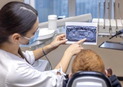
In the world of dental diagnostics, accuracy and early detection of subtle changes within the bone and tissues have become essential. Here at Dalily Medical, Digital Subtraction Radiography (DSR) technology makes a significant difference. This advanced technique relies on comparing two digital images of the same area—then subtracting one from the other to reveal only the new changes, allowing for extremely precise detection of small alterations such as dental caries or bone changes.
DSR is a digital imaging technique that involves taking two X-ray images of the same dental area, such as a tooth or bone, at different times. The first image represents the initial condition, and the second reflects the current state. By digitally subtracting one image from the other, the unchanged parts are canceled out, leaving only the differences visible — such as bone loss, new decay, or changes around a dental implant.
Monitoring Bone Loss or Gain Around Teeth
DSR significantly improves the detection of bone loss compared to traditional X-rays, with the ability to reveal changes as small as 5% in bone density.
Tracking Interproximal Tooth Decay in Children
Studies show DSR measures decay progression as accurately or better than traditional bitewing X-rays, making it a reliable tool for long-term monitoring.
Evaluating Temporomandibular Joint (TMJ) Injuries and Changes
DSR outperforms panoramic imaging in detecting small bony growths (osteophytes) and erosions in the TMJ, with a diagnostic accuracy (ROC) starting at 0.931 versus 0.695 for panoramic X-rays.
Monitoring Bone Healing After Root Canal Treatment
DSR is used to track gradual bone healing around the root of a tooth affected by pulpitis, detecting mineral changes between 90 to 180 days post-treatment.
Assessing Dental Implants
DSR is very useful for monitoring changes in bone density around implants during healing or in cases of peri-implantitis (inflammation around implants).
Early Detection of Degenerative Changes in Jaw Bone
In patients on bisphosphonate drugs (e.g., BRONJ), DSR has been successfully used to quantitatively detect early sclerosis before clinical symptoms appear.
| Advantage | Explanation |
|---|---|
| High Diagnostic Accuracy | Detects very small changes in bone or decay. |
| Reduced Radiation Exposure | Requires fewer images and lower radiation dose. |
| Precise Treatment Monitoring | Ideal for tracking subtle changes over time. |
| Versatile Applications | Useful for decay, implants, TMJ disorders, and more. |
| Quantitative Results | Measures changes with pixel-level or molecular accuracy. |
Precision in Image Capture: Small differences in angle or positioning can affect results.
Specialized Equipment Needed: Maintaining image quality requires dedicated devices for stabilization and alignment.
User Training Required: Dentists and technicians must understand the digital subtraction process to ensure accurate interpretation.
| Imaging Technique | Accuracy | Radiation Exposure | Detection of Small Changes |
|---|---|---|---|
| Traditional X-rays | Good | Medium to High | Very Limited |
| Digital Subtraction Radiography (DSR) | Very High | Low | Highly Effective |
DSR clearly surpasses conventional imaging in early detection, especially for monitoring bone or decay progression with minimal radiation.
Use consistent positioning tools during imaging (e.g., bite blocks or XCP holders).
Keep timing and patient positioning consistent across sessions.
Employ high-quality software for accurate subtraction and image registration.
Train the clinical team on technical standards to avoid errors.
Digital Subtraction Radiography (DSR) represents a significant advancement in diagnosing oral and bone diseases with exceptional accuracy, minimal radiation exposure, and an outstanding ability to track subtle changes like tooth decay, bone density variations, or root canal treatment success. With ongoing improvements in digital tools and user training, DSR is poised to become a standard technique in advanced dental clinics worldwide.


