

Has your doctor recommended a Fluoroscopy (also known as live or moving X-ray)? You might be wondering: what makes it different from a regular X-ray? And are there any risks involved? Don’t worry — in this article from Dalili Medical, we provide you with a comprehensive guide to this important medical imaging technique.
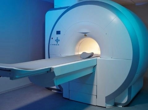
Fluoroscopy is a type of medical imaging that uses continuous X-ray beams to produce real-time moving images of the internal organs. It is often referred to as “live X-ray” because it shows what's happening inside the body in real time—like watching a live video.
Simple analogy: Imagine your doctor watching a live movie of your internal organs while they function—like swallowing food or the heart pumping blood.
| Comparison | Standard X-ray | Fluoroscopy |
|---|---|---|
| Image type | Static (still image) | Dynamic (live video) |
| Duration | One-time snapshot | Continuous imaging for several mins |
| Usage | Diagnosing specific issues | Tracking real-time body functions |
| Radiation level | Lower | Higher (relatively) |
Fluoroscopy is used in many detailed diagnostic and interventional procedures, including:
Such as observing swallowing or the flow of contrast through the esophagus, stomach, and intestines.
Common example: Barium Swallow study.
Like inserting catheters into blood vessels or placing a stent.
In a test called a Cystogram, the bladder is filled with contrast to track its function.
For example, an Arthrogram shows joint movement using contrast dye.
Like cardiac catheterization to examine coronary arteries.
Here’s what typically happens during the test:
Preparation: You may be asked to fast for several hours.
Contrast material: You might ingest or receive an injection of a contrast dye to highlight organs clearly.
Positioning: You will lie on an exam table, and the X-ray machine is adjusted around the area of interest.
Live imaging: The doctor watches live video images and captures snapshots as needed.
Duration: The procedure typically takes between 10 to 30 minutes, depending on the type of test.
The test itself is painless.
However:
You may feel discomfort when swallowing the contrast dye or if a catheter is inserted.
Some people may experience mild nausea due to the contrast agent.
Like all X-ray-based imaging, fluoroscopy uses ionizing radiation. However, it is only recommended when its benefits outweigh the potential risks.
The lowest effective radiation dose is used.
Pregnant women are advised to avoid it unless absolutely necessary.
The rest of the body is shielded with lead aprons when needed.
You can typically resume normal activities immediately, unless you were sedated.
If you took barium contrast, your stool may appear white or light-colored temporarily.
Drink plenty of fluids to help flush the contrast material from your body.
It depends on the type of fluoroscopy being done:
| Type of test | Preparation needed |
|---|---|
| Digestive system | Fasting for 6–8 hours |
| Bladder or kidneys | Drink fluids – Empty your bladder |
| Joints or spine | Usually no special preparation needed |
Pregnant women (unless absolutely necessary for diagnosis).
Those with severe allergies to contrast agents (must inform the doctor).
People with kidney dysfunction (when using certain contrast dyes).
Yes, but only when medically necessary. Doctors usually space out procedures to limit radiation exposure.
Usually not. However, mild sedation may be used in procedures involving children or catheter insertion.
Yes. Doctors can see the live images during the test, and a preliminary report may be provided right after.
Fluoroscopy is a powerful imaging technique that allows doctors to see moving internal structures in real time.
It’s used to diagnose and monitor a wide range of conditions—from the digestive system to the heart.
Despite its use of radiation, it is considered safe when performed under proper medical supervision.
If your doctor recommends a fluoroscopy test, discuss its benefits and potential risks with them to make an informed decision.


