
Uterine abnormalities and effects on reproductive health
Some women suffer from problems related to uterine abnormalities, which are abnormal developments that affect the composition. These distortions can greatly affect a woman's fertility. In addition, uterine abnormalities are divided into several types, where each type is associated with a certain level of risk, especially in the case of desire for pregnancy and childbirth. These abnormalities are considered to be factors that make pregnancy more difficult and require additional effort. Therefore, let's not discuss the problem of uterine abnormalities in a more detailed Dalili Medical, and explain the effects on reproductivehealth.
Does the birth defect in the womb affect the ability to conceive?
In some cases, such as endometrial migration or cervical marriage or uterine marriage, the chances of pregnancy are not increased. Studies have shown that women who suffer from these conditions do not face problems during pregnancy, and can complete the pregnancy period until the ninth month naturally. Also, the degree of fertility of women is not affected by birth defects. In some cases, such as the presence of a uterine barrier, the barrier may be so small that it does not lead to infertility or abortion.
What are the types of birth defects in the womb?
It includes several types of congenital abnormalities in the womb, including al-Hajz-ur-Rahmi, dual-corner, single-corner, and abnormality.
Al-Hajz al-Rahimi: It is considered to be the most common type of congenital malformation, and it is a group of fibrous tissues that divide the uterus partially or completely. Most of the time, this distortion is related to the defect in providing the blood to the area. If the fertilized egg is implanted on the aljaz, the placenta is not able to grow properly, which affects the ability to perform the basic functions of feeding the fetus during pregnancy. This can lead to miscarriage or premature birth before the development of the fetus is complete, which reduces the chances of having a healthy child.
Distortion caused by malformation of the molar tube: This type of distortion occurs as a result of the malformation of the molar tube, which leads to the lack of access to the fallopian tube and cervix, and in some cases, to the vagina as well. This condition is known as MRKH syndrome, and women who suffer from this abnormality can't do it all their lives.
Unicorn uterus: In this type of deformity, a single canal develops only from the double molar canals, which leads to a uterus that is smaller than the normal size, and is curved and elongated in the shape of a banana. However, this distortion does not prevent the existence of the ovaries, as the ovaries continue to produce hormones.
Ovulation is healthy, but the percentage of pregnancy in this case is low, which increases the risk for women, especially in cases of caesarean section, ectopic pregnancy, and premature birth. This type of deformity greatly affects women's fertility.
Urum al-Qarnain, also known as the heart-shaped uterus, arises as a result of the incomplete fusion of the sides of the Mullerian canals. Women who suffer from this deformity are characterized by a uterus with two horns that are smaller in size, where a central barrier separates the two horns, which reduces the space available for the growth of the fetus. This distortion can be classified into two subtypes:
1. Complete uterus: where there is a complete transverse vaginal barrier, it extends to the internal or external opening of the cervix.
2. Al-Urham Zul-Qarnain al-Jaza'i: where the al-Hajz al-Muhabli extends to the inner opening of the womb only.
**Dual uterus:** This is a rare condition characterized by the presence of two types of uterus, and sometimes two types of nipples. This condition may have hereditary roots in the medical history of the family. Most women do not show symptoms before pregnancy, but it increases the risk of miscarriage and increases the possibility of premature birth if pregnancy occurs.
**Al-Rahm al-Maqus:** It is an unconventional shape of the natural uterus, where the ridge that divides the molar canals is small, which gives the uterus an arched shape. This type of malformation rarely leads to infertility, but it may cause a delay in pregnancy. In case of recurrent miscarriage, this condition can be treated surgically.
**Al-Rahm al-Ma'el:** The suffering of women who have this type of uterus is deformation and contraction of the uterus, and the uterus is not visible in ultrasound imaging during the first trimester of pregnancy. There is no evidence that a tilted uterus causes damage or frequent miscarriages. Usually, the inclined uterus returns to its normal position during pregnancy, but in rare cases, it becomes elongated. In this case, surgical intervention is necessary to correct the position of the uterus.
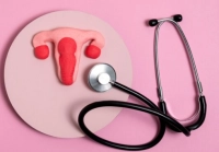
**Description of the female reproductive system** The uterus is an essential part of the female reproductive system, and to explain the deformations of the uterus, it is necessary to understand the components of this system. The female reproductive system consists of the following parts:
**Fallopian canal:** It is a narrow canal connected to the upper part of the uterus, and acts as a tube to transport the egg from the ovary to the uterus. If the egg is fertilized, it reaches the uterus as a fertilized egg ready to implant in the wall of the uterus and develop into an embryo. But if the inoculation is not done, it reaches the uterus, which leads to menstrual bleeding.
**Ovaries:** Both of your small, oval glands are located on the sides of the uterus, and their function is to produce eggs that travel through the fallopian tube to the uterus, in addition to releasing hormones.
**The womb:** It is a hollow organ that rests in the fetus during pregnancy, which lasts for nine months. The uterus is divided into two parts: the cervix, which is the lower part that opens onto the vagina, and the body of the uterus, which is the large cavity that expands easily to fit the different stages of fetal development.
**The vagina**: It is the connecting channel between the cervix and extending to the outside of the body, and it is also known as the birth canal, where it is considered the place where the fetus comes out.
** Causes of uterine deformities **
Uterine deformities usually occur as a result of disturbances in the natural developmental processes of Müller's ligaments during the stage of embryonic development. Müller's canals are the basic structuresThe fusion of the female reproductive system, including the fallopian tube, the uterus, the cervix, and the upper part of the vagina.
Any interruption or deviation during this stage of the height crisis leads to birth defects in the uterus.
**Hereditary factors** Although the exact causes of these developmental disorders may not always be clear, genetic factors play an important role. Mutations in certain genes responsible for the development of the molar canal can lead to the appearance of abnormalities. In addition, the observation of chromosomal abnormalities and family patterns, which indicates the presence of a hereditary element.
**Environmental factors** The influence of environmental factors during pregnancy, such as exposure to substances that cause birth defects, in the development of uterine abnormalities.
Also, the health condition of the mother, inflammation, and the medications you take during pregnancy can play a role in the natural development of Müller's ligaments.
**Congenital defects:** Usually, we have congenital womb defects that are present since birth, where they occur during the formation of the female organs of the child inside the mother's womb. The interpretation of these distortions is due to an error in the formation of the uterus, where there are two tubes known as Muller's canals, and they must be properly combined to form the uterus in the end. And the difference in birth defects based on how these tubes fuse during pregnancy.
**Exposure to harmful radiations:** The exposure of the mother during the pregnancy period to certain types of harmful radiations, which may lead to the deformation of the embryos during the intrauterine development. Therefore, it is necessary to educate pregnant mothers constantly about the dangers of the effects on the health of the fetus.
**Inflammations:** The inflammations inside the uterus can lead to deformities if the treatments are not done in time.
**Some medicines:** Taking some medicines during pregnancy may cause uterine malformations. For example, some mothers who took the drug Diethylstilbestrol (Diethylstilbestrol) known for short as DES, which was used to prevent miscarriages and premature births, exposed their children to birth defects in the womb.
**Classification of uterine abnormalities**
Uterine abnormalities are classified according to the structural characteristics and extent of deviations from the normal structure of the uterus. Prevalence of the American Society of Reproductive Medicine (ASRM) widely accepted classification system, which includes the following categories:
Absence of Müllerian (first letter) **Includes Müller syndrome, also known as Mayer-Rokitansky-Koster-Hauser syndrome (MRKH), complete absence of the uterus and upper third of the vagina. Usually, this condition is diagnosed during adolescence when menstruation does not begin.
**Al-Rahm Zul-Qarn Al-Wahid (Al-Fahta Al-Wahid)** Yhadith al-Rahm Zul-Qarn Al-Wahid is the result of the incomplete development of a single molar canal, which leads to the formation of a single-horned uterus. This anomaly is related to the primitive age, and it may be functional or not.
**Bilateral uterus (the third category)** Distinguishing the condition of the bilateral uterus with the presence of two separated uteruses, where each one has a specific cervix, as a result of failure of fusion of the molar canals. This condition can lead to complications such as recurrent miscarriage and premature birth.
**Zul-Qarnain uterus (fourth grade)** Zul-Qarnain uterus results from the incomplete integration of the molar canals, which leads to a heart-shaped uterus containing two cavities. This anomaly is associated with an increased risk of complications during pregnancy, including cervical insufficiency and fetal malposition.
**Uterus Al-Hajizi (Part 5)** In the case of the divided uterus, the division of the uterus occurs due to a fibrous or muscular barrier, as a result of the lack of complete absorption of the central walls of the molar ducts. This condition is related to the increase in abortion and infertility rates.
**Al-Rahm al-Maqus (Sixth Chapter)** It distinguishes the al-Rahm al-Maqus due to a slight decrease in the bottom of the uterus. This type of uterine abnormalities is considered to be the least severe and often asymptomatic, which leads to a small effect on the reproductive results.
**Anomalies related to diethylstilbestrol (DES) (Seventh category)** Intrauterine exposure to diethylstilbestrol (DES) can lead to structural abnormalities such as a T-shaped uterine cavity. Diethylstilbestrol is a synthetic hormone similar to estrogen. The topic of prescribing for pregnant women between the 1940s and 1970s in the 20th century with the aim of preventing abortion and premature birth, but explaining Later, it causes mood disorders.
**Radiological diagnosis and evaluation**
Diagnosis of uterine abnormalities usually requires a combination of clinical evaluation and imaging studies, and sometimes abdominal examination. Radiological evaluation is considered to be an essential element in accurately determining these distortions and classifications.
**Ultrasound examination** Transvaginal ultrasound is the first imaging method used to analyze the uterus, as it provides accurate images of the uterine cavity and helps in detecting abnormalities such as the divided uterus and the zul-qarnain uterus.
** Magnetic resonance imaging (MRI) ** Magnetic resonance imaging is considered the gold standard for diagnosing uterine abnormalities, thanks to its high ability to differentiate soft tissues and its multi-level capabilities. This test is actively used to distinguish between complex abnormalities and to evaluate associated renal abnormalities.
**Hysterosalpingography (HSG)** includes the operation of dye imaging of the uterus, injection of contrast dye into the cavity of the uterus and fallopian tubes, followed by the operation of x-ray imaging. This technique is used to detect structural abnormalities and is often used to diagnose infertility.
**Symptoms of uterine abnormalities**
**Before marriage:** Some girls may not show any symptoms, while others may suffer from difficulty in menstruation, menstrual irregularity, or pain between now and then.
**After marriage:** After marriage, the main symptom that women notice is repeated miscarriages and difficulty in maintaining pregnancy.
**Pain during sexual intercourse:** Women suffering from gynecological disorders have severe pain during sexual intercourse.
**Diagnosis of uterine abnormalities:** Itam Taaki These symptoms occur during ultrasound imaging or magnetic resonance imaging, according to the instructions of the specialist doctor.
Methods of treating birth defects:
Surgical treatment methods for congenital malformations differ according to the type of malformations, so these malformations are classified into the following categories:
Absence of the vagina. This condition is either part of a syndrome that includes the absence of all other parts of the uterus, or the vagina may be the only missing part while the other parts are intact.
There are several treatment options, whether surgical or non-surgical, aimed at creating a new vagina, which allows the victim to have a normal sexual life.
**Diagnosis:**
- Absence of menstruation.
- The emergence of secondary sexual characteristics naturally.
- The clinical examination of the vagina indicates the absence or presence of a closed small cavity at the end.
- Ultrasound examination sometimes indicates the absence of the uterus and cervix, and this is associated with abnormalities or the absence of the kidney.
- Laparoscopic surgery is performed only in the case of failure of clinical examination and ultrasound in determining the condition, where it is clear that there is no uterus or the presence of a very small and swollen uterus.
**Remedial:**
. **Non-surgical method:** It depends on the use of special dilators of different degrees to create a gap between the iliac opening and the anus. This method requires a long treatment period, lasting from several months to several years, and the surgical method is the best option.
. **Surgical Tariqah:** The main goal of it is to create a vagina for women who want to practice their sexual life naturally, provided that they are psychologically capable of accepting an artificial vagina during the treatment period, which lasts for several months. This is done by using a full fish skin patch after surgery to remove the area, then an artificial vagina is placed inside until the injured area is healed.
**- Al-Jarhariyah:** The main goal of this method is to create a vagina for women who want to practice their sexual life naturally, but are psychologically capable of accepting an artificial vagina during the treatment period, which can last for several months. This is done during the use of a full skin patch, where the area is cut surgically and an artificial vagina is placed inside until the injured area heals.
**- Al-Rahm Dhul-Qarn Al-Wahed:** This distortion occurs as a result of the growth of one of the canals of Malbin, either completely or partially. In this case, the womb will be zu qarn al-wahid, accompanied by the presence of a closed and closed qurn.
**Surgical treatment:** There is no surgical treatment for the single-cornered uterus except in one case, which is the presence of endometrium inside the closed corner of the uterus, which leads to the accumulation of blood inside this closed part of the uterus. In this case, the patient needs to undergo a laparoscopic operation to remove the uterine part of the uterus.
**- Double uterus:** This condition occurs as a result of the complete or partial non-union of the Malerian ducts, which leads to the existence of two uteruses and a single cervix.
**Surgical treatment:** Surgical treatment varies according to the degree of deformity, as the condition may include:
- Marriage of the uterus with the cervix and obstruction in the vagina due to the membrane, which requires surgical intervention to remove the vaginal septum. Abdominal angiography can also be performed at the same time to remove any adhesions in the pelvis or to treat other problems such as endometriosis.
- Marriage of the uterus and cervix with the presence of a single open vagina, and this does not require any surgical intervention.
I am glad that these cases do not cause major problems, as studies have shown that most cases of pregnancy can last until the ninth month, and the fertility level of these women is good.
**- Urmah Dhu al-Qarnain:** This hadith is a distortion as a result of complete non-union of the Malerian canal, where the lower part of the cervix is fused, while the upper part is not fused. As a result, there are two cavities in the uterus and the septum that separates them, and it is distinguished by the presence of an external protrusion that separates the upper parts of the uterus. The difference between the depth of this opening and the size of the wall is different from one patient to another, depending on the degree of connection between the canals of Malrin.
It is not possible to rely on the color x-ray image of the uterus to diagnose this condition, as it does not give a clear image of the outer wall of the uterus. Three-dimensional ultrasound examination is considered the best way to diagnose these conditions.
**Surgical treatment:** It includes surgical treatment of the unification of the upper part of the womb, and in the presence of the cervix, it is necessary for the unification of the two parts. You should avoid performing vaginal endoscopy in these cases, as it carries risks for the patient and may lead to a hole in the uterus.
**Al-Hajj al-Rahmi:** Hadith of Al-Hajj al-Rahmi as a result of non-destruction of Al-Hajj al-Wasati, which separates the canals of Malrin when they join them. This barrier is located in the middle of the uterus and is characterized by the density of muscle fibers in the composition, in addition to the lack of arteries and blood vessels. Al-Hajj can be complete or small and partial, which does not cause problems such as infertility or repeated miscarriages.
**Surgical treatment:** The preferred surgical treatment in these cases is the endometriosis, where the cervix is expanded and the endometriosis is inserted. It is preferable to perform a laparoscopic procedure to avoid any problems during the operation, such as perforation of the uterine wall. It is also recommended to place a uterine tube during the operation to prevent adhesions. There is a controversy surrounding the position of the IUD after the operation, as some point out that the IUD causes inflammation leading to adhesions inside the uterus. And in some cases, the patient may need to undergo a second operation after three months, if there is a part of the barrier.
**Al-Rahm Al-Munbaaj** It is distinguished by a slight bulge in the upper wall of the uterus. This type of deformity does not require any surgical intervention, as it is not related to pregnancy or fertility problems compared to the normal state. From the above, we can conclude that the diagnosis and treatment of uterine deformities has become easier, faster and more accurate at the moment, thanks to the availability of detection devices. And modern treatment.

