
Published on: 2026-11-19 | Written by: Dr. Ahmed Adel Shaheen, Consultant Cardiologist
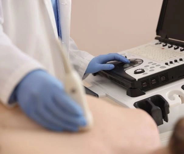
Transthoracic Echocardiogram (TTE) and Transesophageal Echocardiogram (TEE) represent two of the most vital and accurate non-invasive and semi-invasive diagnostic imaging tests in modern cardiology. Both rely on safe ultrasound waves to generate live, moving images of the heart, its valves, chambers, and connected major blood vessels. Standard transthoracic echo (TTE) is used as a primary, rapid examination to evaluate heart muscle efficiency and valves from outside the chest wall, whereas transesophageal echo (TEE) provides an ultra-clear, close-up view of the heart from behind via the esophagus, given the esophagus's direct proximity to the left atrium and heart valves without interference from bones or lungs. Performing and interpreting these examinations requires high medical precision to ensure an accurate diagnosis that guides the treatment plan with maximum safety.
Transthoracic and transesophageal echocardiograms aim to evaluate the pumping efficiency of the heart muscle, examine heart wall motion, detect valve regurgitation or stenosis, and identify any blood clots or congenital defects with extreme precision prior to making therapeutic or surgical decisions.
Dr. Ahmed Adel Shaheen clarifies that the integration between standard transthoracic echo and transesophageal echo gives the cardiologist a comprehensive, high-resolution picture of the heart's functional and anatomical status, noting that transesophageal echo is critically necessary when a case requires closer inspection of the valves, ruling out blood clots inside the left atrium, or evaluating the efficiency of prosthetic valves and complex congenital defects.
Evaluating Heart Muscle Efficiency and Ejection Fraction: Measuring the left ventricle's ability to pump blood to the rest of the body's organs and monitoring the heart's response to treatment.
Inspecting Heart Valves with High Precision: Detecting cases of narrowing (stenosis) or leakage (regurgitation) in the mitral, aortic, tricuspid, or pulmonary valves and accurately determining their severity.
Ruling Out Cardiac Clots and Valve Infections: Early detection of any blood clots in the left atrium or inflammatory vegetations on the valves, particularly through transesophageal echocardiography.
Diagnosing Congenital Defects and Aortic Aneurysms: Examining the septa between heart chambers and the ascending aorta with high precision, and guiding catheter-based or surgical interventions.
Cardiac ultrasound imaging has witnessed major technological advancements, with devices now equipped with color Doppler technologies and three-dimensional/four-dimensional (3D/4D) imaging that highlight the finest anatomical details and valve movements in real time.
Dr. Ahmed Adel Shaheen points out that relying on 3D/4D echocardiography devices, Tissue Doppler techniques, and Myocardial Strain Imaging allows for detecting early changes in heart muscle function even before any obvious symptoms appear or before a drop in the traditional ejection fraction occurs.
Color and Pulsed Doppler Ultrasound: Measuring the speed and direction of blood flow across heart valves and chambers to assess the severity of regurgitation or stenosis.
Three-Dimensional and Four-Dimensional Echocardiography (3D/4D Echo): Providing live, clear three-dimensional models of heart valves and their details to facilitate planning for catheter or surgical interventions.
Myocardial Strain Imaging (Speckle Tracking): Detecting hidden or early weakness in heart muscle fibers with high accuracy in patients with hypertension, diabetes, or those receiving chemotherapy.
Fine Transesophageal Echo Probe (TEE Probe): A thin, flexible tube equipped with high-frequency ultrasound transducers passed into the esophagus to obtain ultra-clear images of the heart.
Preparation and procedural steps differ between the non-invasive standard echo (TTE) and the transesophageal echo (TEE), which requires fasting and local throat anesthesia; both follow a structured medical protocol ensuring patient comfort and safety.
Dr. Ahmed Adel Shaheen advises patients to adhere to fasting instructions for at least 6 hours before undergoing a transesophageal echo and to inform the physician of any swallowing difficulties, esophageal issues, or drug allergies, emphasizing that a standard transthoracic echo requires no fasting or prior preparation and is performed effortlessly within a few minutes.
Patient Preparation and Explaining Examination Steps: Explaining the test to the patient and reassuring them, alongside fasting and numbing the throat with a local anesthetic or mild sedative in the case of a transesophageal echo.
Lying in the Appropriate Position: Lying mostly on the left side to bring the heart closer to the chest wall in a standard echo or closer to the esophagus in a transesophageal echo.
Applying the Transducer or Passing the Esophageal Probe: Placing water-based gel and the ultrasound transducer on the chest for a standard echo, or gently inserting the esophageal probe after numbing the throat for a transesophageal echo.
Capturing Images and Doppler Measurements: Recording comprehensive clips of the heart chambers, valves, and blood flow velocity with high accuracy.
Concluding the Exam and Post-Procedure Monitoring: Wiping off the gel and resuming activities immediately after a standard echo, whereas waiting for the throat anesthetic to wear off before eating or drinking is required after a transesophageal echo.
Adhering to simple post-examination instructions—especially following a transesophageal echo—alongside adopting a heart-healthy lifestyle plays an important role in stabilizing the patient's condition and executing the treatment plan effectively.
Dr. Ahmed Adel Shaheen stresses that waiting until the local throat anesthetic wears off completely before eating or drinking hot beverages after a transesophageal echo prevents any choking, emphasizing that adhering to pharmacological therapy and controlling cardiovascular risk factors represent the foundation for protecting the heart muscle and valves.
Waiting Until Normal Throat Sensation Returns After TEE: Avoiding eating or drinking for one to two hours until the local anesthetic effect completely disappears.
Resting on the Day of the Transesophageal Echo: Refraining from driving on the same day if a sedative was used, and avoiding very hot drinks during the first few hours to prevent throat irritation.
Adhering to Prescribed Heart and Blood Pressure Medications: Continuing to take cardiac treatments according to doctor instructions based on the examination results.
Maintaining a Balanced, Heart-Healthy Diet: Reducing salt, saturated fats, and sugars while increasing intake of vegetables, fruits, and whole grains.
Engaging in Appropriate Physical Activity Under Medical Supervision: Following the level of movement and exercise determined by the cardiologist according to heart muscle and valve efficiency.
Do Not Delay an Echocardiogram When Cardiac Symptoms Exist: Consult a physician early upon experiencing unusual shortness of breath, palpitations, swelling in the feet, or chest pain.
Inform the Physician of Any Swallowing Difficulties or Esophageal Conditions Before TEE: To ensure selecting the safest timing and method for the patient.
Commit to Periodic Follow-Ups of Heart Valves and Muscle Efficiency: Undergoing echocardiography at scheduled intervals to track any changes early.
Maintain Ongoing Follow-Ups with Your Specialist:
Dr. Ahmed Adel Shaheen emphasizes in his concluding guidance that standard transthoracic echocardiography and transesophageal echocardiography represent a decisive diagnostic pillar in cardiac patient care, confirming that early, accurate diagnosis, adherence to medical instructions, and regular follow-ups are the true guarantees for protecting the heart muscle and valves and enjoying a safe, active, and healthy life.