
Published on: 2026-11-17 | Written by: Dr. Ahmed Mohamed Khamis, Consultant Orthopedic
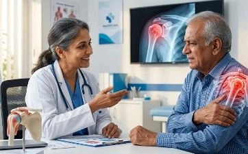
Bone deformities are medical issues that go far beyond affecting the external appearance of the body; they cause an imbalance in how weight and loads are distributed across joints and the spine, leading over time to chronic pain, difficulty walking, and early joint osteoarthritis. With major advancements in modern orthopedic surgery, bone deformity correction procedures have become precise and safe solutions to restore bones to their natural axis and recover the patient's full motor function.
Dr. Ahmed Mohamed Khamis, Consultant of Orthopedic Surgery, Fractures, Joint Replacement, Arthroscopy, and Bone Deformity Correction at Alexandria University and Member of the American Academy of Orthopaedic Surgeons (AAOS), provides the latest surgical techniques for diagnosing and treating various bone deformities in children and adults according to the highest international medical standards.
Bone deformities refer to any abnormal deviation in a bone's shape, length, angular alignment, or rotation compared to healthy anatomical placement. This deformity may be present from birth (congenital), develop during growth stages in childhood and adolescence, or result from past injuries and fractures in adults.
The bone deformities managed by Dr. Ahmed Mohamed Khamis include:
Bow Legs / Knock Knees: Whether curving outward (bowed legs) or inward (knock knees), these are among the most common deformities in the lower limbs.
Short or Unequal Limb Length: A clear length discrepancy between legs or arms due to congenital defects or injuries affecting growth plates.
Malunion Deformities: Occurring when an old fracture heals at a skewed angle or with rotation, impeding limb movement and causing persistent pain.
Foot and Ankle Deformities: Such as clubfoot, severe flatfoot (bony flatfoot), and toe alignment abnormalities.
Spinal Deformities: Such as scoliosis (lateral curvature) or severe kyphosis.
Upper Extremity Deformities: Such as forearm or elbow misalignment resulting from childhood fractures.
Various factors lead to the development of bone deformities, most notably:
Congenital and Genetic Factors: Issues occurring during embryonic development affecting bone growth or causing the absence of a part.
Metabolic Diseases and Vitamin Deficiencies: Such as osteomalacia and rickets resulting from vitamin D and calcium deficiencies, rendering bones soft and prone to bending under body weight.
Growth Plate Injuries: Children experiencing fractures or infections in bone growth plates may experience halted growth on one side and continued growth on the other, causing progressive angular deviation.
Complications from Old Fractures: Neglecting fracture treatment or failing to immobilize them in proper alignment leads to healing at a deformed angle.
Chronic Bone and Joint Diseases: Such as advanced osteoarthritis causing wear on one side of the knee joint and secondary bowing.
Chronic Bacterial Bone Infections (Osteomyelitis): Which can affect bone structure and alignment.
Dr. Ahmed Mohamed Khamis emphasizes that early intervention to correct bone deformities protects patients from severe long-term complications, most notably:
Early Joint Osteoarthritis: Limb axis deviation concentrates total body weight onto a small portion of the knee or ankle cartilage, causing rapid wear.
Lower Back and Pelvic Pain: Resulting from body imbalance and limping during walking, especially in cases of limb length discrepancy.
Difficulty Moving and Performing Daily Activities: Experiencing rapid fatigue and an inability to walk long distances or exercise.
Psychological and Social Impact: Particularly in children and adolescents due to altered gait or limb appearance.
A successful correction plan relies on precise pre-surgical measurements, with medical evaluations including:
Comprehensive Clinical Examination: To assess limb axis during standing and walking, measure joint range of motion, and examine muscle and nerve strength.
Long-Leg Panoramic X-Ray (Scanogram / Long-Leg X-Ray): The primary scan used to measure deviation angles with extreme precision, determine the deformity center, and quantify leg length discrepancies.
3D CT Scan: Used in complex deformities or those involving rotational bone twists to map an exact geometric layout of the bone pre-operatively.
Laboratory Tests: To verify calcium, vitamin D levels, endocrine function, and bone metabolic activity prior to surgical intervention.
Treatment approaches vary based on patient age, deformity type, and severity, including:
In early stages for children, treatment may involve addressing vitamin deficiencies and utilizing medical splints.
Guided Growth Technique: A very minor surgical procedure placing a tiny plate across one side of the growth plate to temporarily slow growth on the healthy side, allowing the other side to grow and naturally correct leg alignment without fracturing the bone.
When is it used? In cases of pronounced bowing or deformities resulting from malunited fractures in adolescents and adults.
How is it performed? The surgeon makes a precisely calculated surgical cut in the deformed bone to immediately restore normal alignment, then secures it using advanced locking plates and screws or intramedullary nails.
When is it used? In complex multi-level deformities, cases of bone shortening and limb length discrepancies, or deformities accompanied by prior infections.
Concept: Utilizing precise external fixation devices (such as the Ilizarov apparatus or modern circular fixators) allowing gradual correction and bone lengthening at a rate of 1 millimeter per day, stimulating the body to generate strong new natural bone tissue in the resulting gap.
Deformity correction surgeries demand a blend of geometric precision and deep surgical expertise, which Dr. Ahmed Mohamed Khamis provides:
Specialized Academic Expertise: Consultant of Orthopedic Surgery, Fractures, Joint Replacement, Arthroscopy, and Bone Deformity Correction at Alexandria University, and Member of the American Academy of Orthopaedic Surgeons (AAOS).
Precise Pre-Surgical Geometric Planning: Accurately calculating correction angles to ensure the mechanical axis of the leg returns to its optimal position, protecting joints from osteoarthritis.
Selecting the Most Suitable Method per Patient: Carefully balancing fast internal fixation and gradual external fixation to achieve the highest levels of patient safety and comfort.
Ongoing Follow-Up and Rehabilitation: Continuous supervision of bone healing and physical therapy programs to recover muscle strength and full flexibility.
Adhering to Weight-Bearing Protocols: The doctor determines the appropriate timing to initiate partial and then full weight-bearing on the leg using crutches according to the type of fixation used.
Physical Therapy Sessions: Play a pivotal role in maintaining joint flexibility and strengthening muscles surrounding the corrected bone.
Bone-Healing Nutritional Support: Emphasizing proteins, calcium, and vitamin D, while strictly avoiding smoking as it slows bone healing.
Follow-Up X-Rays: Periodic imaging to verify complete bone healing and stability of corrected angles.