Published on: 2026-10-29 | Written by: Dr Amir Ramadan Jumaa Professor of Ophthalmology
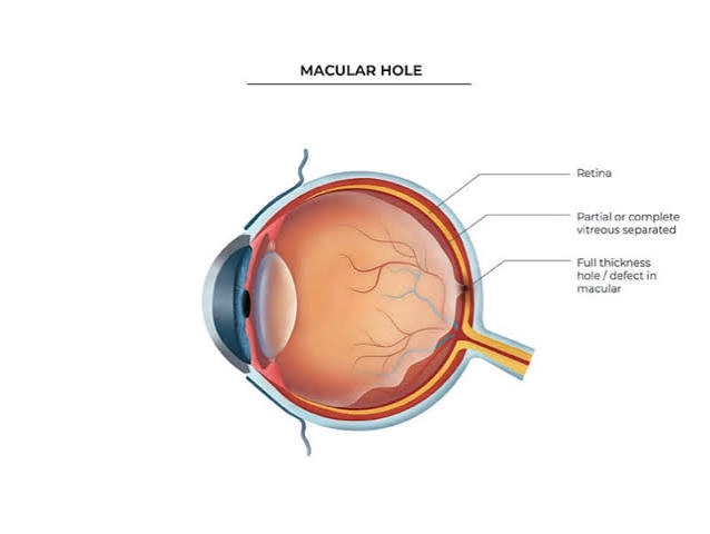
Advanced fundus conditions such as retinal detachment and macular holes are critical medical emergencies in ophthalmology that directly threaten vision by affecting the sensitive neural layers responsible for transmitting visual images to the brain. Retinal detachment occurs when fluid leaks through a retinal tear, separating the retina from its underlying nourishing vascular layer. Conversely, a macular hole represents a microscopic defect in the macular center, which is responsible for sharp, detailed central vision and reading ability. Managing these conditions requires urgent, high-precision microscopic surgical intervention to stabilize ocular tissues and save vision from permanent loss.
Therapeutic plans for these conditions aim to reposition the retinal layers back to their normal anatomical location, seal tears and holes, and evacuate subretinal fluid accumulation to ensure the recovery of visual function.
Dr. Amir Ramadan Jomaa clarifies that treating retinal detachment and macular holes represents a precise challenge in vitreoretinal micro-surgery, noting that the speed of surgical intervention as soon as symptoms appear is the decisive factor for successful visual restoration and for protecting sensitive neural cells from irreversible damage.
Reattaching the Detached Retina: Re-fusing delicate neural layers to the vascular wall of the eye to ensure proper blood supply and nourishment.
Closing the Macular Hole Precisely: Re-approximating the edges of the hole in the macular center to enhance reading capability and fine-detail perception.
Relieving Vitreous Traction: Evacuating fibrotic gel fibers that pull on the retina and cause tears and holes to widen.
Preserving the Visual Field: Protecting the patient from complete vision loss and restoring central and peripheral visual efficiency.
Fundus surgery has witnessed a monumental technological evolution thanks to micro-incision vitrectomy techniques, specialized surgical stains, and modern intraocular tamponade agents.
Dr. Amir Ramadan Jomaa points out that relying on ultra-fine micro-vitrectomy techniques and delicate staining agents to peel microscopic retinal membranes has dramatically raised the success rates of closing macular holes and treating detachment safely and effectively.
Micro-Incision Vitrectomy: Suctioning out the traction-inducing gel and replacing it with supportive medical fluids or gas.
Medical Gas or Silicone Oil Injection: Placing tamponade agents inside the eye to secure the edges of the macular hole and retina throughout the healing period.
Advanced Surgical Stains: Meticulously identifying membrane layers during macular hole surgery to avoid any cellular trauma.
Complementary Laser or Cryotherapy: Creating therapeutic chorioretinal scars around retinal tears to prevent recurrent detachment.
Retinal detachment and macular hole surgeries follow a strict, structured medical protocol starting with comprehensive radiological diagnosis and concluding with adherence to home recovery positioning.
Dr. Amir Ramadan Jomaa advises strictly maintaining the prescribed head positioning following macular hole surgery (such as keeping the head facing downward for several days), emphasizing that this posture is the key to successful fusion of the hole's edges and its return to normal structure.
Precise Diagnosis and Optical Coherence Tomography (OCT): Evaluating macular hole size and the extent of retinal detachment with absolute digital accuracy.
Surgical Preparation and Local or General Anesthesia: Selecting a safe anesthetic protocol tailored to the patient's overall health status.
Microscopic Surgical Intervention: Performing vitrectomy, membrane peeling, and injecting gas or silicone tamponade.
Recovery and Postural Adherence: Monitoring gradual gas absorption and central vision improvement through periodic follow-up visits.
Long-Term Follow-Up: Evaluating retinal stability and intraocular pressure to prevent any future relapses.
The post-surgical phase serves as the primary foundation for ensuring retinal stability and macular hole healing without adverse complications.
Dr. Amir Ramadan Jomaa stresses that completely avoiding air travel while a gas bubble is present, strictly adhering to therapeutic eye drops, and refraining from physical and mental exertion represent golden rules for ensuring ocular safety.
Strict Adherence to Head Positioning (Face-Down): Maintaining the recommended head angle to ensure the gas bubble presses directly against the macular center.
Refraining from Air Travel Temporarily: Completely avoiding flying as long as gas remains inside the eye to prevent bubble expansion and dangerous IOP spikes.
Consistent Use of Antibiotic Eye Drops: Applying prescribed medications meticulously to prevent post-operative bacterial infections.
Avoiding Eye Rubbing or Pressure: Protecting the eye by wearing a protective medical shield during sleep and rest.
Avoiding Heavy Lifting: Abstaining from strenuous physical effort that could elevate intraocular pressure and compromise healing tissues.
Immediate Action Upon Emerging Symptoms: Never ignoring the sudden appearance of flashing lights, a dark curtain over vision, or distorted letters and lines.
Meticulous Control Over Chronic Diseases: Regulating blood sugar and systemic blood pressure levels to protect delicate retinal vessels.
Full Adherence to Follow-Up Schedules: Assessing gradual gas absorption and ensuring the safety of the macular center.
Maintain Ongoing Follow-Ups with Your Specialist:
Dr. Amir Ramadan Jomaa emphasizes in his concluding guidance that the astonishing progress in retinal detachment and macular hole surgery techniques has yielded remarkable success rates and restored sight to millions. He confirms that early awareness, immediate surgical intervention, and strict adherence to recovery instructions are the true shields for strong and sustainable visual health.