

Published on: 2026-09-08 | Written by: Dr. Ahmed El-Kashki, Lecturer of Neurosurgery
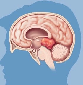
Brain and Skull Base Tumors
Brain and skull base tumors represent one of the most delicate and complex medical specialties in neurosurgery. Managing them requires deep knowledge of complex neuroanatomy, along with the utilization of the latest surgical and interventional techniques to ensure safe tumor resection while protecting the patient's vital functions.
What are Brain and Skull Base Tumors?
Brain Tumors: Abnormal cell growths within the brain tissue. They can be benign (slow-growing and non-invasive) or malignant (rapidly growing and invading surrounding tissues).
Skull Base Tumors: Tumors that grow in the complex lower region of the skull, which separates the brain from the face and neck. This area houses major blood vessels and critical cranial nerves responsible for vision, hearing, movement, and balance.
Most Common Types of Tumors
Meningiomas: Arise from the membranes surrounding the brain. They are among the most common brain and skull base tumors and are often benign.
Pituitary Adenomas: Grow in the pituitary gland located at the base of the skull, affecting hormonal balance and vision.
Acoustic Neuromas: Originate on the nerve responsible for hearing and balance.
Gliomas: Develop from supportive cells in the brain and vary in their severity.
Symptoms Requiring Medical Consultation
Symptoms vary depending on the size and location of the tumor within the brain or skull base, including:
Persistent or worsening headaches, especially early in the morning.
New-onset seizures or convulsions.
Vision changes (such as double vision or blurred vision) or problems with hearing and balance.
Weakness or numbness on one side of the face or limbs.
Difficulty speaking or swallowing, as well as changes in personality or memory.
Advanced Diagnostic Methods
Accurate diagnosis relies on a series of high-precision imaging and functional tests:
Magnetic Resonance Imaging (MRI): The primary test for determining the tumor's location, size, and relationship to surrounding tissues.
Computed Tomography (CT Scan): Used to evaluate the bones and structural anatomy of the skull base.
CT Angiography and Diagnostic Cerebral Angiography: Used to assess the tumor's blood supply and its proximity to major blood vessels.
Modern Treatment Options
The treatment plan depends on the type, size, and location of the tumor, and includes:
1. Advanced Microsurgery
Utilizing surgical microscopes and 3D neuro-navigation systems to reach and resect the tumor with maximum precision without harming vital brain centers.
2. Endoscopic Skull Base Surgery
Resecting certain skull base tumors and pituitary tumors through the nose using an endoscope, avoiding large surgical incisions in the skull and shortening recovery time.
3. Radiation Therapy and Stereotactic Radiosurgery
Directing highly precise radiation beams to destroy tumor cells or shrink the tumor without surgery. Used for small tumors or those in surgically inaccessible locations.
4. Periodic Follow-Up and Monitoring
For small, benign, symptom-free tumors, regular monitoring through periodic MRI scans may be sufficient.
Early diagnosis and relying on specialized medical expertise in brain and skull base surgery are the most crucial steps to achieving optimal treatment outcomes and restoring the patient's quality of life.