

Published on: 2026-08-22 | Written by: Dr. Samar El-Sayed Madani Catheterization Consultant
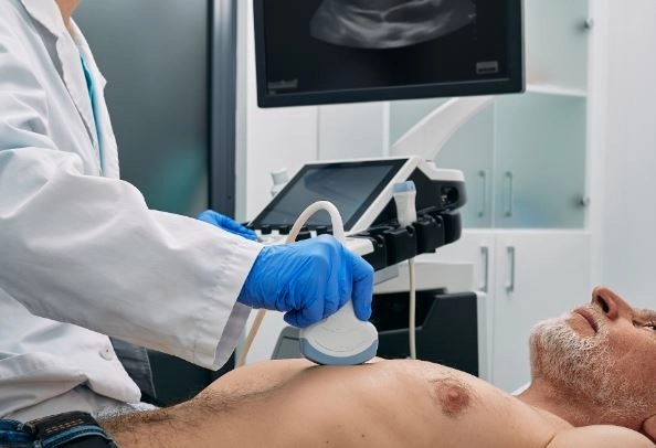
Dr. Samar El Sayed Madany explains that echocardiography, or an echocardiogram, is one of the most important non-invasive tests used to evaluate the structure and function of the heart, heart muscle movement, valves, and blood flow, as it uses sound waves to create moving images of the heart while it is beating and can support the diagnosis and monitoring of many cardiac conditions.
Echocardiography is a medical diagnostic procedure that uses high-frequency sound waves to produce clear, moving images of the heart, helping physicians evaluate the size and shape of the heart, wall thickness, heart muscle movement, pumping function, and valve performance.
The importance of echocardiography is that it helps assess heart muscle strength, contraction, and movement, and may reveal changes in cardiac function that require follow-up or additional testing, with results interpreted alongside the patient’s symptoms, medical history, clinical examination, and other diagnostic tests.
Dr. Samar El Sayed Madany confirms that evaluating heart pumping function is an important part of echocardiography, as the examination allows physicians to assess heart muscle performance and the movement of different chambers, while measurements such as the ejection fraction may be used as part of the overall assessment of cardiac function.
Heart valve evaluation allows echocardiography to assess how the valves open and close and how blood flows through them, helping physicians identify certain valve conditions such as narrowing or regurgitation and monitor how these problems affect heart function over time.
Color Doppler is a technique that can be used with echocardiography to evaluate the direction and speed of blood flow through the heart and across the valves, providing additional information when assessing certain valve disorders or abnormalities in blood flow.
Heart failure assessment relies in part on echocardiography to evaluate heart muscle movement, chamber size, pumping function, and valve performance, while the results are considered together with clinical examination, ECG findings, laboratory tests, and other investigations when necessary.
Dr. Samar El Sayed Madany explains that follow-up echocardiography may be important for certain cardiac patients to monitor changes in heart muscle or valve function and compare current findings with previous examinations, while the timing of repeat echocardiography depends on the patient’s condition, disease severity, symptoms, and previous results.
Heart valve assessment highlights the importance of echocardiography in determining the severity of valve narrowing or regurgitation and its effect on the heart, helping physicians decide whether the patient requires regular monitoring, additional testing, or medical intervention according to the overall clinical assessment.
Heart chamber evaluation allows physicians to assess chamber size, wall thickness, and movement, providing valuable information when symptoms or medical conditions may affect the structure or function of the heart and helping monitor changes over time.
Evaluation of a heart murmur may lead a physician to request an echocardiogram to determine whether there is a heart valve or structural abnormality, as the examination provides direct images of heart movement and valve function and can help determine whether further monitoring or testing is required.
Evaluation of cardiac symptoms may result in an echocardiogram being recommended when a patient experiences shortness of breath, unexplained chest discomfort, palpitations, leg swelling, or symptoms associated with heart or valve function, with the appropriate examination selected according to the patient’s medical history and clinical findings.
An echocardiogram is generally a simple, painless, and non-invasive examination, during which the patient lies down while a healthcare professional applies ultrasound gel and moves a transducer over the chest to obtain images of the heart, without using X-rays or ionizing radiation during a standard echocardiogram.
A standard echocardiogram generally requires little or no special preparation, while instructions may vary depending on the type of echocardiographic examination, as specialized procedures such as a transesophageal echocardiogram require specific preparation provided by the medical team.
Different types of echocardiography include transthoracic echocardiography, transesophageal echocardiography, stress echocardiography, and three-dimensional echocardiography, with the physician selecting the most appropriate type according to the information needed and the patient’s condition and symptoms.
Echocardiography can help identify certain blood clots inside the heart chambers and abnormal changes around the valves or the surrounding structures of the heart, although additional diagnostic tests may sometimes be required to confirm a diagnosis or determine its underlying cause.
Dr. Samar El Sayed Madany confirms that performing echocardiography and color Doppler under the supervision of a qualified specialist helps ensure accurate assessment of the images and measurements and allows the findings to be interpreted alongside the patient’s symptoms and medical history.
Comprehensive cardiac assessment shows that echocardiography is an important tool for evaluating the heart muscle, chambers, valves, pumping function, and blood flow, but it cannot diagnose every heart condition on its own, and physicians may need to combine echocardiography with ECG, laboratory tests, stress testing, imaging, or other investigations depending on the patient’s condition.
Regular cardiac follow-up is important for patients with known heart conditions, as scheduled examinations can help monitor heart and valve function and detect changes at an appropriate stage, allowing the physician to adjust the treatment or follow-up plan when necessary.