

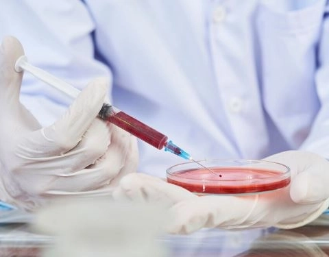
Surgical removal of a tumor is an important step in the treatment journey. However, surgery alone is not always enough to determine the exact nature of the tumor or how successfully it has been removed. This is why tumor tissue analysis after surgical removal is an essential step in identifying the characteristics of the tumor cells, determining whether the tumor is benign or malignant, establishing its grade, and assessing whether it has been completely removed.The analysis results also help doctors evaluate the risk of tumor recurrence and determine whether the patient needs additional treatments, such as chemotherapy or radiation therapy, or whether regular follow-up is sufficient.So, what are the different types of tests performed on a tumor after surgical removal? How are they conducted? When are the results available? And how can patients understand a pathology report? We will explore all these questions in this article from Dalili Medical.
Tumor analysis after surgical removal is a medical examination performed on tissue samples removed from a patient's body during surgery. Its primary purpose is to determine the nature of the tumor and identify whether it contains cancerous cells. This examination is known as a pathological examination or anatomical pathology analysis.
The examination is performed by specialized physicians known as pathologists, using various techniques, including microscopic tissue examination, immunohistochemical staining, and molecular and genetic testing when necessary. The results help determine the tumor type, grade, and characteristics, as well as assess whether it has been completely removed surgically, contributing to the development of an appropriate treatment plan for each patient.
Tumor tissue analysis is performed after surgical removal to accurately determine the nature of the tumor and establish whether it is benign or malignant. It also helps identify the cell type, tumor grade, and other important characteristics.
Additionally, examining the surgical margins helps determine whether the edges of the removed tissue are free of tumor cells and whether further treatment or regular follow-up may be necessary.
Yes, a tumor can be malignant even if surgery successfully removes it completely. Complete surgical removal does not necessarily mean that the tumor is benign; rather, it indicates that the visible tumor tissue has been removed within the boundaries of the excised specimen.
The need for additional treatment depends on the tumor type, stage, grade, and biological characteristics, along with the results of other medical examinations.
Yes. Pathological examination is one of the most important medical methods used to determine the nature of a tumor. A pathologist examines tissue samples and cells under a microscope to identify the changes that distinguish benign tumors from malignant ones.
In some cases, additional tests, such as immunohistochemical staining or molecular testing, may be necessary to reach a more accurate diagnosis.
Yes, tumor analysis results may be inconclusive in certain cases due to similarities in cellular characteristics or difficulties in accurately identifying the tumor type through microscopic examination alone.
In such situations, the doctor may request additional tests, such as immunohistochemistry (IHC), molecular testing, or genetic analysis, to help establish an accurate diagnosis.
No. A delay in receiving tumor analysis results does not necessarily indicate that the tumor is malignant. The delay may be due to the need for additional examinations, specialized immunohistochemical staining, or a review of the tissue sample by a specialist pathologist before issuing the final report.
Examining the surgical margins helps assess whether the removed tissue is free of tumor cells at its edges.
If the results show cancer cells at one of the margins, this may indicate the possibility of residual tumor cells at the surgical site. The doctor must then evaluate the case to determine whether additional surgery or other treatments are needed, depending on the tumor type and characteristics.
Not necessarily. The treatment plan varies depending on the tumor type, grade, stage, and pathological examination results.
Surgery alone may be sufficient to treat certain tumors, while other types may require additional treatments, such as chemotherapy, radiation therapy, or targeted therapy, to reduce the risk of recurrence or control any remaining cancer cells.
Yes. Some tumors may recur even after complete surgical removal. The likelihood of recurrence varies depending on the tumor type, biological characteristics, grade, and stage, as well as the patient's response to treatment.
Therefore, regular medical follow-up and periodic examinations are essential for detecting any signs of tumor recurrence at an early stage.
Tumor tissue analysis alone cannot accurately determine the patient's chances of recovery. However, it provides important information about the tumor's nature, type, grade, and characteristics. When combined with clinical examinations, imaging studies, and other medical tests, these results help doctors assess the patient's condition and predict the possible course of the disease.
Yes. A tumor sample can be reexamined, or previously preserved tissue slides and specimens can be reviewed, particularly when the initial results are unclear, a second medical opinion is needed, or additional immunohistochemical and molecular tests are required to confirm the diagnosis.
Yes. The results differ depending on the nature of the cells, their characteristics, and their growth patterns.
Benign tumors generally consist of cells that do not show signs of cancerous invasion into surrounding tissues. In contrast, malignant tumors may exhibit abnormal changes in cell appearance and organization, as well as the ability to invade nearby tissues and spread to other parts of the body.
Patients may need additional tests after receiving their pathology results, depending on the tumor type, its characteristics, and the surgical findings.
These tests may include blood tests, computed tomography (CT) scans, magnetic resonance imaging (MRI), or other imaging procedures. Their purpose is to evaluate the surgical site, detect any remaining tumor tissue or signs of disease spread, and determine the most appropriate treatment and follow-up plan.
The results of a fibroid tumor analysis are often available within 3 to 7 business days at many laboratories. However, the turnaround time may vary depending on the sample size, tissue characteristics, and examination procedures involved.
In some cases, the laboratory may need to perform immunohistochemical staining or additional tests to accurately identify the nature of the cells, which may delay the results by several additional days.
The types of tests and examinations a patient may need after surgical tumor removal vary depending on the tumor's nature, location, size, and the purpose of the surgery.
These tests help identify the tumor type and characteristics, assess the patient's overall health, monitor surgical outcomes, and detect any remaining tumor tissue or potential signs of recurrence.
The main examinations are divided into three categories:
Tumor tissue examinations are among the most important diagnostic procedures performed after surgery. They help identify the nature of the removed tissues and cells, determine whether the tumor is benign or malignant, and provide important information about its characteristics.
The main tests include:
Histopathological Examination: This test involves examining a tumor tissue sample under a microscope to identify the cell type and tissue structure and detect characteristics that help distinguish benign tumors from malignant ones.
Immunohistochemistry (IHC): This technique is used to detect specific proteins and markers within cells. It helps identify the exact type and origin of a tumor and may contribute to selecting the most appropriate treatment.
Molecular and Genetic Testing: These tests aim to detect specific genetic mutations and alterations within tumor cells. Their results may help classify certain tumors and determine whether targeted therapies are suitable.
Surgical Margin Examination: This examination determines whether tumor cells are present at the edges of the removed tissue. It helps assess the completeness of surgical removal and identify whether additional treatment may be necessary in certain cases.
Doctors may request several blood tests to assess the patient's overall health, monitor recovery after surgery, and help detect certain potential complications.
The most important tests include:
Complete Blood Count (CBC): This test evaluates hemoglobin levels, white blood cells, and platelets. It can help detect anemia and certain signs of infection.
Liver and Kidney Function Tests: These tests assess how well the liver and kidneys are functioning, particularly when planning to use certain medications or additional treatments.
Blood Clotting Tests (PT, PTT, and INR): These tests evaluate the blood's ability to clot properly. They may be requested, particularly in patients with bleeding disorders or those taking anticoagulant medications.
Tumor Markers: These are specific substances measured in the blood. They may be used in certain types of cancer to monitor treatment response or help detect possible disease recurrence. However, tumor markers alone are not definitive evidence of cancer.
Patients may need certain imaging tests to monitor the surgical site, evaluate surrounding tissues, and detect any remaining tumor tissue or signs of disease spread. The most common imaging examinations include:
Computed Tomography (CT Scan): Helps visualize the area from which the tumor was removed, assess surrounding tissues, and detect certain abnormal changes or fluid collections after surgery.
Magnetic Resonance Imaging (MRI): Provides detailed images of tissues and organs and is used to monitor certain tumors, particularly brain tumors, to detect any remaining tumor tissue or changes at the surgical site.
Ultrasound: Used to monitor the surgical site and examine tissues and internal organs in certain cases, such as breast, thyroid, and abdominal tumors.
Positron Emission Tomography–Computed Tomography (PET-CT): Combines CT imaging with an assessment of the metabolic activity of tissues. It may be used for certain types of malignant tumors to evaluate disease spread or monitor treatment response.
Tumor analysis after surgical removal begins by sending the surgical specimen to a pathology laboratory, where it undergoes a series of procedures and laboratory examinations to determine the nature of the tissues and cells, establish whether the tumor is benign or malignant, and assess its characteristics and the completeness of surgical removal.
The main steps involved in this process include:
After the tumor is surgically removed, the specimen is placed in a specialized container, usually containing an appropriate concentration of formalin to preserve the tissues and prevent decomposition.
It is then sent to the pathology laboratory along with the patient's medical information, details about the tumor's location, and information about the surgical procedure.
The pathologist performs a visual examination of the specimen with the naked eye to document its characteristics and identify the areas that require microscopic examination.
This examination includes:
Measuring the tumor's size and determining its dimensions and weight.
Describing the tumor's shape, color, and consistency.
Assessing the surrounding tissues.
Determining its relationship to nearby tissues and organs.
Examining the surgical specimen margins and identifying their locations.
The specimen undergoes several laboratory procedures, beginning with tissue fixation and processing using appropriate chemicals.
The tissues are then embedded in paraffin wax blocks and cut into extremely thin sections using a specialized instrument called a microtome. These sections are placed on glass slides in preparation for staining and microscopic examination.
The tissue sections are stained using special dyes, the most common of which is Hematoxylin and Eosin (H&E).
The pathologist then examines the slides under a microscope to study the appearance, arrangement, and characteristics of the cells and identify any abnormal changes that may help determine the nature and type of the tumor.
In some cases, conventional microscopic examination may not be sufficient to establish an accurate diagnosis. Therefore, the doctor may request specialized tests, including:
Immunohistochemistry (IHC): Used to detect specific proteins and markers within cells, helping identify the tumor type and its origin.
Molecular and Genetic Testing: Used to detect specific genetic mutations or alterations that may help classify the tumor and determine the most appropriate treatment.
Surgical Margin Examination: Determines whether tumor cells are present at the edges of the removed tissue and helps assess the completeness of surgical removal from a pathological perspective.
After completing the required examinations, the pathologist prepares a final report containing the main diagnostic findings, including:
The tumor type and nature.
Whether the tumor is benign or malignant.
The tumor grade, when applicable.
The status of the surgical margins.
Any additional tissue or cellular characteristics that may affect the evaluation of the patient's condition and treatment plan.
After receiving the report, the patient should discuss the findings with the treating physician or specialist surgeon to review the diagnosis, assess the success of surgical removal, and determine the next steps.
In some cases, regular follow-up may be sufficient. However, other cases may require additional examinations or complementary treatments, such as chemotherapy, radiation therapy, or targeted therapy, depending on the tumor type, its characteristics, and the results of other medical tests.
Tumor analysis results after surgical removal reveal the nature of the tissues and cells removed during surgery. They help determine the tumor type, its characteristics, and the completeness of surgical removal. They also contribute to assessing the risk of tumor recurrence and determining whether additional treatment or regular follow-up is necessary.
The meaning of the results varies depending on the tumor type and its characteristics. Below is an explanation of the most common terms that may appear in a pathology report:
Benign Tumor: This means that the tumor is non-cancerous and does not invade surrounding tissues or spread to distant organs. Surgical removal may be sufficient to treat it in many cases, depending on the tumor type and location.
Malignant Tumor: This indicates the presence of cancerous cells that have the ability to invade surrounding tissues and may spread to other parts of the body, depending on the cancer type, stage, and characteristics.
Negative Surgical Margins: This means that no tumor cells were detected at the edges of the examined surgical specimen, suggesting that the tumor was completely removed within the boundaries of the excised tissue. However, this does not necessarily rule out the presence of tumor cells elsewhere in the body.
Positive Surgical Margins: This means that tumor cells are present at one or more edges of the removed specimen, which may indicate the possibility of residual tumor cells at the surgical site. This may require an assessment of whether additional surgery or adjuvant treatment is necessary, depending on the tumor type and the patient's overall health.
Tumor Grade: This describes how different tumor cells are from normal cells and provides an indication of the tumor's growth pattern and potential behavior. Some tumors are classified into low, intermediate, or high grades, with the significance of each grade varying according to the tumor type.
No Evidence of Malignancy: This means that the examination did not reveal malignant cells in the analyzed sample. The findings may be consistent with a benign tumor or non-cancerous tissue, depending on the specimen description and the other details included in the report.
Cellular Atypia: This refers to abnormalities in the shape, size, or structure of cells compared with normal cells. These changes do not necessarily indicate cancer, as their significance depends on their severity, the tissue type, and the other examination findings.
Immunohistochemistry Markers: These markers are used to help identify the type and origin of tumor cells and distinguish between different types of tumors. Their results may also contribute to selecting the most appropriate treatment for certain tumors.
Cell Proliferation Index (Ki-67): This indicates the approximate percentage of tumor cells undergoing proliferation and division. A higher percentage may help assess the activity of certain tumors; however, it cannot be used alone to determine the level of risk or predict the course of the disease.
The time required to analyze a tumor sample after surgical removal varies depending on the specimen type, tumor characteristics, required examinations, and whether additional tests are needed to reach an accurate diagnosis.
In general, the approximate turnaround times for different examinations are as follows:
Routine Histopathological Examination (Histopathology): Usually takes 3 to 7 business days. However, the timeframe may be longer depending on the specimen size, case complexity, and whether additional tissue sections need to be examined.
Immunohistochemistry (IHC): May require several additional days, typically ranging from 3 to 10 business days, depending on the number of stains required and their availability in the laboratory.
Molecular and Genetic Testing: May take 1 to 3 weeks or longer, depending on the type of test, the techniques used, and the laboratory performing the examination.
Intraoperative Frozen Section Examination: Results are usually available within 15 to 30 minutes. This examination is performed in certain cases during surgery to help the surgeon make immediate decisions regarding the extent of tumor removal. However, it is considered a preliminary examination and does not always replace the final histopathological analysis.
Several factors may increase or decrease the time needed to issue tumor analysis results. The most important include:
The size of the sample and the amount of tissue removed.
The type of tumor and the complexity of its cellular structure.
Whether additional immunohistochemical stains or molecular and genetic tests are required.
Whether the sample needs to be reviewed by a specialized pathology consultant.
The laboratory’s workload and the availability of the necessary equipment and technologies.
A tumor pathology report (Pathology Report) contains important medical information that helps determine the nature of the tumor, the characteristics of its cells, and whether it has been completely removed surgically.
The report may seem complicated because it contains specialized medical terminology. However, its main sections can be understood by becoming familiar with the meanings of the following key terms:
This section identifies the type of tissue sent to the laboratory for examination and the organ or area from which it was removed, such as the brain, breast, thyroid gland, or another part of the body. It may also describe how the sample was obtained, whether through complete tumor removal or a biopsy involving only part of the tumor.
This is one of the most important sections of the report. It presents the conclusion reached by the pathologist after examining the tissue and may include one of the following terms:
Benign: A benign tumor, meaning it does not contain cancerous cells and does not invade nearby tissues or spread to distant organs.
Malignant: A malignant tumor, meaning it contains cancerous cells that can invade surrounding tissues and may spread to other parts of the body.
Borderline: A tumor with borderline characteristics that do not fully meet the criteria for either benign or malignant tumors. Its interpretation depends on the tumor type and its specific features.
No Evidence of Malignancy: No malignant cells were identified in the analyzed sample.
This section indicates the tumor’s dimensions in centimeters or millimeters and helps assess its size. Tumor size may be an important factor in determining the stage of certain cancers, along with other factors such as invasion into nearby tissues or involvement of lymph nodes.
Tumor grade describes how different the tumor cells are from normal cells and provides an indication of the tumor’s potential behavior and activity. It may be classified as follows:
Low Grade: Indicates that the cells generally differ less from normal cells and tend to grow more slowly.
Intermediate Grade: Indicates characteristics that fall between low-grade and high-grade tumors.
High Grade: Usually indicates cells that differ significantly from normal cells and may grow more rapidly and behave more aggressively.
The method used to determine tumor grade and its significance vary depending on the type of tumor.
This section indicates whether tumor cells are present at the edges of the removed tissue. It helps assess whether the tumor has been completely removed within the boundaries of the examined specimen. The report may include the following terms:
Negative Margins: No tumor cells are present at the examined edges of the specimen.
Positive Margins: Tumor cells are present at one or more edges of the specimen, which may indicate that some tumor cells remain at the surgical site.
Close Margins: The distance between the tumor and the edge of the specimen is small. The significance of this finding depends on the measured distance stated in the report and the type of tumor.
This section indicates whether cancer cells have entered the blood vessels or lymphatic vessels within the tissue. The presence of lymphovascular invasion is one of the factors that may affect the assessment of the likelihood of tumor spread and the selection of a treatment plan, depending on the cancer type and stage.
This term refers to the presence of tumor cells surrounding nerves or extending within their sheaths. It may be an important feature when assessing certain types of cancer, as it can be associated with tumor behavior and the likelihood of local recurrence or spread.
If immunohistochemical staining is required, the report may include the results of several protein markers that help identify the tumor type and origin and distinguish between different cell types. Some of the most important markers include:
Ki-67: A marker used to estimate the rate of cell proliferation within the tumor.
CD20 and CD3: Markers that help identify specific types of lymphocytes and may be used in diagnosing certain lymphatic system tumors.
ER, PR, and HER2: Markers used to classify certain breast tumors. Their results also help determine the most appropriate treatment options.
The report may include the results of molecular or genetic tests designed to detect specific mutations or changes within tumor cells. These findings help complete the diagnosis, identify the characteristics of certain tumors, and may contribute to selecting targeted therapies or determining whether a patient is likely to benefit from particular treatments.
This section contains the pathologist’s overall findings. It may clarify whether the diagnosis is definitive or indicate the need for additional tests, such as immunohistochemical staining or molecular testing, to complete the evaluation of the sample.
The report may also include notes that help the treating physician interpret the findings in the context of the patient’s medical condition and other test results.
When reviewing a tumor pathology report after surgical removal, several key sections can help you understand the tumor’s nature and characteristics, assess the completeness of its removal, and identify factors that may influence the treatment and follow-up plan.
The following table summarizes the most important sections and what each one indicates:
| Report Section | What Does It Indicate? |
|---|---|
| Final Diagnosis | The final diagnosis, identifying the nature and type of the tumor and whether it is benign or malignant. |
| Tumor Size | The tumor’s dimensions and overall size, which may help determine the stage of certain cancers. |
| Tumor Grade | The tumor’s grade and how much its cells differ from normal cells, helping assess its potential behavior. |
| Surgical Margins | The status of the edges of the removed specimen and whether they are free of or contain tumor cells. |
| Lymphovascular Invasion | Whether tumor cells are present inside blood vessels or lymphatic vessels. |
| Perineural Invasion | Whether tumor cells are present around nerves or within their sheaths. |
| Immunohistochemistry (IHC) | Cellular characteristics and immune markers that help identify the tumor type and origin. |
| Molecular Testing | Genetic and molecular changes within tumor cells, which may help complete the diagnosis and guide the selection of certain treatments. |
Note: Not all of these sections necessarily appear in every pathology report. The information and tests required vary according to the tumor type and the affected organ. Results should also be interpreted collectively rather than relying on a single finding in isolation.