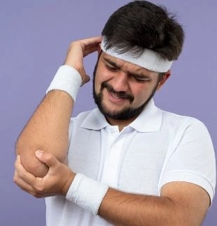
An olecranon fracture, also known as a fracture of the elbow tip, is one of the common injuries affecting the elbow joint. It often occurs as a result of a direct fall on the arm or a strong impact. Although the elbow may appear to be a simple joint in terms of movement, it is actually one of the most important joints for performing daily activities such as bending, extending, and carrying objects.When this type of fracture occurs, the patient may experience sudden severe pain, noticeable swelling, and difficulty moving the arm, making prompt medical intervention essential to avoid complications such as stiffness or reduced elbow mobility.In this article we will discuss olecranon fractures in detail, including their causes, symptoms, diagnostic methods, treatment options, and the most important tips to ensure full recovery and restore normal elbow function.
What is an Olecranon Fracture (Elbow Tip Fracture)?
An olecranon fracture is a break in the prominent bony part of the ulna at the elbow, which forms the back tip of the elbow joint. This type of fracture commonly occurs due to a direct fall onto the elbow or a strong blow to the area.
Is an Olecranon Fracture Serious?
✔ In most cases, it is not considered dangerous.
However, the severity increases in the following situations:
- If the fracture is significantly displaced
- If the fracture is comminuted (broken into several pieces)
- If the fracture extends into the joint
- If it is associated with nerve injury or elbow dislocation
Does an Olecranon Fracture Always Require Surgery?
❌ No, not always.
✔ Some simple, non-displaced fractures can be treated with casting or immobilization alone.
However, in many cases — especially displaced or comminuted fractures — surgery is necessary to restore the bones to their normal position and stabilize them properly.
How Long Does It Take for an Olecranon Fracture to Heal?
The healing time depends on the type of fracture and the treatment method, but generally:
- Initial bone healing takes about 6 to 8 weeks
- Full recovery and restoration of strength and movement may take 3 months or longer
Can the Elbow Return to Normal After the Fracture?
✔ In most cases, the elbow can return to normal or near-normal function.
This depends on several important factors, including:
- Early diagnosis and treatment
- Commitment to physical therapy and rehabilitation
- Avoiding complications such as joint stiffness or limited motion
When Can I Start Moving My Elbow After the Fracture?
The timing of elbow movement is determined by the doctor and depends on the type of fracture and treatment method:
- In some surgical cases, early movement may begin after about two weeks
- In simpler cases, movement usually starts after cast removal
⚠️ Random or premature movement should be avoided because it may negatively affect fracture healing.
Do Metal Plates or Screws Need to Be Removed Later?
✔ In some cases, yes — especially if:
- The patient experiences pain or irritation from the hardware
- The patient is a child or a highly active young adult
❌ However, not all patients need hardware removal, and many live comfortably with the implants permanently.
Why Can’t I Fully Straighten My Elbow After the Fracture?
Difficulty straightening the elbow after an olecranon fracture is common initially and may result from:
- Pain caused by the injury
- Swelling around the joint
- The effect on the triceps muscle attached to the olecranon
However, if the problem persists for a long time, medical evaluation is recommended.
Can Elbow Stiffness Occur After the Fracture?
✔ Yes, elbow stiffness is one of the most common complications after this type of fracture.
However, the risk can be greatly reduced by:
- Starting physical therapy at the appropriate time
- Gradually moving the joint once approved by the doctor
When Can I Return to Work or Sports?
- Light work: usually after 4 to 6 weeks
- Heavy activities or sports: after 2 to 3 months, depending on the doctor’s assessment
Recovery time depends on the severity of the fracture and the speed of healing.
Is Pain After Casting or Surgical Fixation Normal?
✔ Yes, some pain is normal in the beginning.
It should gradually improve over time.
⚠️ However, if the pain becomes worse or remains severe, medical attention is needed immediately.
Types of Olecranon Fractures
Olecranon fractures are classified into several types depending on the fracture pattern, degree of bone fragmentation, and involvement of the joint.
1. Stable / Non-Displaced Fracture
- The bone is broken but remains in its normal position
- No obvious separation between bone fragments
- Often treated with casting or immobilization without surgery
- Pain may be present, but visible deformity is usually minimal
2. Displaced Fracture
- The bone fragments are separated from each other
- There may be visible deformity or irregularity around the elbow
- Surgical fixation with plates, screws, or wires is often required
3. Comminuted Fracture
- The bone breaks into multiple small fragments
- Commonly caused by severe trauma or accidents
- Requires precise surgical reconstruction and fixation
4. Intra-Articular Fracture
- The fracture extends into the elbow joint surface itself
- May affect joint movement in the long term
- Requires accurate treatment to reduce the risk of future arthritis or joint stiffness
5. Fracture-Dislocation
- Involves both an olecranon fracture and elbow dislocation
- Considered a complex injury that often requires urgent surgical treatment
- Significantly affects joint stability
Causes of Olecranon Fracture
1. Direct Fall on the Elbow
One of the most common causes occurs when a person falls directly onto the back of the elbow while trying to protect themselves.
2. Falling on an Outstretched Hand
When a person falls with the hand extended, the force travels from the hand through the forearm to the elbow, which may result in an olecranon fracture.
3. Strong Direct Trauma
Such as road traffic accidents or violent blows to the elbow during work or sports activities.
4. Sports Injuries
Certain contact sports may lead to this type of fracture due to falls or strong impact, including:
5. Osteoporosis
Especially in older adults, where weakened bones make even a minor fall capable of causing an olecranon fracture.
6. Forceful Triceps Muscle Contraction
In rare cases, a sudden and powerful contraction of the triceps muscle may pull on the olecranon and cause a fracture, particularly when the bones are weak.
Symptoms of Olecranon Fracture (Elbow Tip Fracture)
An olecranon fracture usually presents with several clear symptoms that often appear immediately after the injury, including:
1. Severe Elbow Pain
- The pain is usually sharp and sudden after a fall or direct impact
- It becomes significantly worse when trying to move the arm or straighten the elbow
2. Noticeable Swelling
- Rapid swelling develops around the elbow joint
- The swelling may increase during the first few hours after the injury
3. Bruising or Skin Discoloration
- Blue or reddish discoloration may appear around the elbow
- The bruising may sometimes extend to the forearm
4. Inability to Straighten the Arm
- The patient may have difficulty or inability to fully extend the elbow
- This occurs because the fracture affects the olecranon and the function of the triceps muscle
5. Deformity or Abnormal Prominence
- The elbow may appear misshapen
- There may be a visible depression or abnormal contour behind the joint
6. Sensation of Clicking or Instability
- The patient may feel that the joint is unstable
- A grinding or clicking sound may be noticed during movement
7. Pain When Pressing on the Elbow
- Even light touch or pressure on the elbow area may cause severe pain
Diagnosis of Olecranon Fracture (Elbow Tip Fracture)
Diagnosis of an olecranon fracture begins with a clinical examination and is confirmed using imaging studies to accurately determine the type and severity of the fracture.
First: Clinical Examination
The doctor starts by taking the medical history and examining the elbow. Important findings may include:
- Severe pain when pressing on the elbow bone
- Significant swelling around the elbow joint
- Difficulty or inability to straighten the arm
- Bruising or skin discoloration
- Possible deformity or abnormal prominence
- Examination of sensation and movement in the hand and fingers to ensure the nerves are intact
Second: X-Ray Examination
X-rays are the primary diagnostic tool because they:
- Clearly show the presence of the fracture
- Identify the fracture type (non-displaced, displaced, or comminuted)
- Determine whether the fracture extends into the joint
- Are usually taken from multiple views (front and side views)
Third: CT Scan
A CT scan is used in more complex cases, such as:
- Comminuted fractures
- Fractures extending into the joint
- Cases not clearly visible on regular X-rays
This imaging helps the doctor plan treatment or surgery more accurately.
Fourth: Evaluation of Nerves and Blood Vessels
In some cases, doctors also check for:
- Integrity of the ulnar nerve
- Adequate blood flow to the hand and arm
- Absence of numbness or weakness in the fingers
Complications of Olecranon Fracture (Elbow Tip Fracture)
Although many olecranon fractures heal well, complications may occur if treatment is delayed or performed incorrectly.
First: Complications of Delayed or Improper Treatment
1. Weakness or Loss of Elbow Extension
The olecranon is closely related to the function of the triceps muscle. Improper healing may lead to permanent weakness in straightening the arm.
2. Elbow Joint Stiffness
The elbow is highly sensitive to movement restriction. Inadequate rehabilitation may result in stiffness and difficulty bending or extending the joint.
3. Nonunion
In some cases, the bone may fail to heal completely, causing chronic pain and instability of the elbow joint.
Second: Functional and Structural Complications
4. Malunion
The fracture heals in an abnormal position, which may cause deformity or limited movement.
5. Reduced Arm Strength
Even after healing, some patients may notice decreased arm strength or difficulty carrying objects.
Third: Neurological Complications
6. Ulnar Nerve Injury
Compression or irritation of the ulnar nerve may cause:
- Numbness in the ring and little fingers
- Weakness in the hand muscles
Fourth: Surgical Complications (If Surgery Is Performed)
7. Infection or Inflammation
Infection may develop around the plates or wires and may require medical treatment.
8. Irritation or Pain from Metal Hardware
Some patients may feel discomfort from the implanted hardware, and in certain cases, removal may be needed later.
Fifth: Elbow Joint Arthritis
If the fracture extends into the joint, cartilage damage may develop over time, leading to:
- Chronic pain
- Difficulty moving the elbow
Medical Treatment of Olecranon Fracture
The primary treatment for an olecranon fracture is immobilization or surgery. However, medications play an important role in pain relief, reducing inflammation, and supporting recovery.
First: Painkillers
Pain management is especially important during the first days after injury.
1. Paracetamol (Acetaminophen)
- Used to relieve mild to moderate pain
- Considered relatively safe for most patients
- Reduces pain sensation without strong anti-inflammatory effects
2. Nonsteroidal Anti-Inflammatory Drugs (NSAIDs)
Examples include:
- Ibuprofen
- Diclofenac
- Naproxen
✔ These medications help to:
- Reduce pain
- Decrease swelling
- Relieve inflammation around the fracture site
⚠️ They should be used cautiously in patients with:
- Stomach disorders
- Kidney problems
Long-term use without medical supervision is not recommended.
Second: Medications to Reduce Inflammation and Swelling
In some cases, additional medications may be prescribed to reduce inflammation.
- Corticosteroids are rarely used in olecranon fractures.
Third: Antibiotics
Antibiotics are used only in specific situations, such as:
✔ Open fractures
When the skin is injured and the bone is exposed to air, antibiotics are used to prevent infection.
✔ After surgery
To reduce the risk of infection around plates, screws, or wires.
Fourth: Bone-Healing Support Supplements
Although not essential, some supplements may help support healing, such as:
- Vitamin D
- Calcium
- Bone-strengthening supplements
✔ These may improve bone healing
✔ Especially beneficial for older adults or patients with osteoporosis
Fifth: Blood Thinners (In Some Cases)
Doctors may prescribe mild blood thinners in situations such as:
- Prolonged immobilization
- After surgery
⚠️ These medications should only be used under direct medical supervision.
Surgical Treatment of Olecranon Fracture – Types and Procedures
Surgical treatment is often necessary for displaced, comminuted, or intra-articular olecranon fractures to restore normal bone alignment and elbow function.
First: Tension Band Wiring
✔ Suitable for:
- Simple non-comminuted fractures
- Displaced fractures without severe fragmentation
Procedure
- The bone is repositioned into its normal alignment
- Metal wires are inserted into the bone
- A tension band wire is used to hold the fragments together
- The pulling force of the triceps muscle is converted into compression that promotes bone healing
⭐ Advantages
- Relatively simple surgery
- Faster recovery
- Lower cost
⚠️ Disadvantages
- Possible irritation of tissues beneath the skin
- Hardware removal may be needed after healing
Second: Plate and Screw Fixation
✔ Suitable for:
- Severely displaced fractures
- Comminuted fractures
- Fractures extending into the joint
Procedure
- Surgical opening of the elbow area
- Precise repositioning of bone fragments
- Fixation using a metal plate
- Stabilization with strong screws
⭐ Advantages
- Very strong fixation
- Ideal for complex fractures
- Allows earlier movement after surgery
⚠️ Disadvantages
- Larger surgery compared to wiring
- Possible discomfort from the hardware
- Plate removal may be required in some cases
Third: Partial Olecranon Excision
✔ Suitable for:
- Elderly patients
- Severely comminuted fractures that cannot be repaired
Procedure
- Removal of the fractured bone fragment
- Reattachment of the triceps tendon to preserve elbow function
⭐ Advantages
- Useful for difficult and complex cases
- Provides rapid pain relief
⚠️ Disadvantages
- May reduce elbow extension strength
- Usually not suitable for younger patients
Fourth: External Fixation (Rarely Used)
✔ Suitable for:
- Severe open fractures
- Contaminated wounds or major skin injuries
- Complex injuries around the elbow
Procedure
- Metal supports are attached outside the arm
- Pins connect the external frame to the bone
- Stabilization is achieved externally without major internal surgery
⭐ Advantages
- Suitable for severe injuries
- Reduces the risk of deep infection
⚠️ Disadvantages
- Uncomfortable appearance for the patient
- Requires careful maintenance and regular follow-up
Fifth: Arthroscopic-Assisted Fixation (Modern Technique)
✔ Suitable for:
- Intra-articular fractures
- Cases requiring highly precise fixation
Procedure
- A small camera is inserted into the elbow joint
- Bone fragments are repositioned accurately
- The fracture is fixed using specialized internal instruments
⭐ Advantages
- Very high precision
- Less damage to surrounding tissues
- Better functional outcomes in some cases
Tips for Managing an Olecranon Fracture
Following proper instructions after an olecranon fracture is very important to reduce pain, promote bone healing, and prevent complications.
First: During the First 48 Hours After Injury
✔ Immobilize the Arm
- Keep the arm as still as possible
- Use a splint or brace as instructed by the doctor
- Avoid moving or testing the elbow
✔ Elevate the Arm
- Keep the elbow elevated above heart level
- This helps reduce swelling and pain
✔ Apply Cold Compresses
- Apply ice for 15–20 minutes every 2–3 hours
- Helps reduce swelling and inflammation
Second: Follow the Treatment Plan
- Take pain medications as prescribed
- Do not stop treatment without consulting the doctor
- After surgery, complete the prescribed antibiotics course
Third: Things to Avoid
❌ Random elbow movements
❌ Lifting or carrying weight with the injured arm
❌ Using the arm to push yourself while standing or sitting
❌ Forcing the elbow to straighten
Fourth: Cast and Wound Care
If Wearing a Cast
- Keep the cast completely dry
- Do not insert objects inside the cast
- Seek medical advice if severe pain or unusual swelling occurs
After Surgery
- Clean the wound according to medical instructions
- Watch for signs of infection such as redness, warmth, or discharge
Fifth: Follow-Up Care
- Perform X-rays at scheduled appointments
- Attend all follow-up visits
- Assess elbow movement once healing begins
Sixth: Physical Therapy (After Doctor Approval)
- Starts gradually and not too early
- Includes gentle exercises for elbow bending and extension
- The main goal is to prevent joint stiffness
- Should be supervised by a specialist
Seventh: Nutrition to Support Bone Healing
- Calcium-rich foods such as milk, cheese, and yogurt
- Vitamin D from sunlight or supplements
- Protein-rich foods such as meat, eggs, and legumes
- Drink adequate amounts of water
⚠️ Warning Signs That Require Immediate Medical Attention
- Increasing pain instead of improvement
- Numbness in the fingers
- Severe swelling or discoloration of the hand
- Fever or discharge from the surgical wound