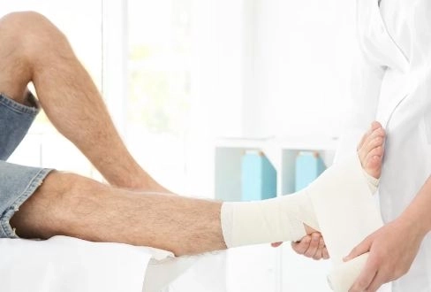
A calcaneus fracture (heel bone fracture) is one of the most painful injuries and has a direct impact on a person’s ability to move and walk normally. The heel bone is the largest bone in the foot and is responsible for supporting body weight during standing and movement.This type of fracture usually occurs due to falls from a height, severe accidents, or sudden strong pressure on the foot. Although a calcaneus fracture can be serious in some cases, early diagnosis and proper treatment make a significant difference in speeding up recovery and reducing complications.Treatment varies depending on the type and severity of the fracture, ranging from conservative treatment using a cast or splint to surgical intervention in more complex cases. After healing, rehabilitation exercises are very important to restore normal movement and regain walking ability safely.In this Delili Medical article, we will explore everything you need to know about heel bone fractures, including their causes, symptoms, diagnostic methods, and treatment options, as well as exercises that help you walk again without pain and in a safe way.
What is the heel bone and why is it so important? (The foundation)
The calcaneus (heel bone) can be compared to the foundation of a building. It supports the entire body weight during standing, walking, and running, and also acts as the main shock absorber when the foot hits the ground.
Its importance is not limited to that. The heel bone is a key part of the subtalar joint, which is responsible for the side-to-side movement of the foot. This allows the foot to adapt to uneven surfaces during movement.
For this reason, calcaneus fractures are not only about bone healing; they also require precise reconstruction of the joint surface. Failure to restore proper joint alignment may lead to complications such as:
- Chronic pain
- Osteoarthritis (joint degeneration)
- Difficulty and abnormal walking
❓ 1) Is a heel bone fracture serious?
Yes, a calcaneus fracture is considered a significant and relatively serious injury, especially if it is:
- Intra-articular (involving the joint), or
- Comminuted (broken into multiple fragments)
This is because it can greatly affect walking and mobility for a long period. However, most cases improve well with early diagnosis and proper treatment.
❓ 2) Can you walk on a heel bone fracture?
❌ No, it is strictly forbidden to bear weight or walk on the injured foot in cases of a calcaneus fracture.
Walking in this condition may lead to:
- Worsening of the fracture
- Bone deformity
- Delayed healing
Therefore, doctors usually recommend using:
- Crutches
- A cast or splint depending on medical instructions
❓ 3) How long does it take to heal a heel bone fracture?
Healing time usually ranges from:
- 6 to 12 weeks in simple cases
- 3 to 6 months in severe cases or after surgery
It is important to note that rehabilitation and physical therapy may continue even after the bone has healed to fully restore normal movement.
❓ 5) Does a heel bone fracture leave permanent effects?
In some cases, a calcaneus fracture may leave lasting effects such as:
- Chronic foot pain
- Stiffness in the ankle or heel joint
- Difficulty walking normally
However, early diagnosis and proper treatment significantly reduce the risk of these complications.
❓ 6) When can I return to normal walking?
You can return to walking gradually:
- After proper bone healing
- With adherence to a physiotherapy program
Full recovery usually occurs within:
3 to 6 months, depending on the severity of the fracture and treatment type.
❓ 8) Can the fracture happen again in the same place?
Yes, but it is rare.
The risk may increase in cases such as:
- Weak bones (e.g., osteoporosis)
- Another severe fall or injury
❓ 10) Is a cast enough to treat all heel bone fractures?
❌ No, not in all cases.
A cast is used only for:
- Simple, non-displaced fractures
In more severe cases such as:
- Displaced fractures
- Comminuted fractures
- Intra-articular fractures
Surgery may be necessary for better healing and functional outcomes.
Types of calcaneus (heel bone) fractures
1) Simple (non-displaced) fracture
This is a crack or fracture in the bone without movement of the bone fragments.
- The bone remains in its normal position
- No visible deformity of the heel occurs
- Usually treated with a cast and rest
- Tends to heal relatively quickly
2) Displaced fracture
In this type, the bone fragments move out of their normal position.
- The shape of the heel changes
- It causes noticeable pain and swelling
- It usually requires more intensive medical treatment
- Surgical fixation may be necessary
3) Comminuted fracture
This is considered one of the most serious types of fractures.
- The bone breaks into several small pieces
- It often results from severe trauma or falling from a height
- In most cases, surgery is required
- Healing time is relatively longer
4) Intra-articular fracture
The fracture extends into the heel joint itself.
- It directly affects foot movement
- It may lead to stiffness or arthritis after healing
- Requires precise diagnosis and close follow-up
- Surgery may be needed to restore the joint surface
5) Extra-articular fracture
The fracture does not involve the joint.
- Less severe than intra-articular fractures
- Does not directly affect the joint surface
- Often responds well to conservative treatment (such as casting)
6) Stress fracture
A small crack caused by repeated pressure on the bone.
- Develops gradually, not suddenly
- Common in athletes or with overuse
- Causes pain that worsens with activity
- Requires rest and reducing weight-bearing on the foot
Causes of calcaneus (heel bone) fracture
1) Falling from a height
This is the most common cause.
- Falling from stairs
- Falling from a roof or elevated surface
- The heel absorbs a strong impact when hitting the ground
2) Severe trauma (road accidents)
Common causes include:
- Car accidents
- Motorcycle accidents
- Direct force or strong twisting of the foot
3) Sports injuries
Usually occur in high-impact sports involving jumping or hard landings, such as:
- Gymnastics
- Parkour
- Landing incorrectly on the foot
4) Direct heel impact
Such as:
- A heavy object falling directly on the heel
- A strong blow to the bottom of the foot
5) Repetitive stress (stress fractures – rare)
Caused by:
- Repeated pressure on the bone over time
- Common in athletes or physically active individuals
6) Osteoporosis (especially in older adults)
In this condition, the bone becomes weak and brittle, making it more prone to fractures even from minor injuries due to:
- Calcium deficiency
- Osteoporosis
Symptoms of calcaneus (heel bone) fracture
1) Severe heel pain
- Sudden, sharp pain immediately after injury
- Worsens when standing or putting weight on the foot
- May persist even at rest
2) Swelling
- Rapid swelling in the heel after injury
- May spread to the foot and ankle
3) Bruising and skin discoloration
- Blue or purple bruises around the heel
- May extend to the sides and sole of the foot
4) Difficulty or inability to walk
- Unable to bear weight on the affected foot
- Walking becomes very difficult or impossible due to pain
5) Heel deformity (in severe cases)
- The heel may appear flattened or abnormal
- The foot may look shorter than normal
6) Pain on touch
- Even light pressure causes significant pain
- Increased sensitivity in the injured area
7) Limited ankle movement
- Difficulty moving the foot up or down
- Stiffness around the heel joint
- Reduced flexibility
Diagnosis of calcaneus (heel bone) fracture
1) Clinical examination
The first step includes:
- Taking medical history (fall, accident, or strong impact)
- Careful physical examination of the foot and heel
- Observing signs such as:
- Severe swelling
- Bruising
- Sharp pain on pressure
- Difficulty or inability to walk
2) X-ray
- Usually the first imaging test requested
- Helps detect fractures or cracks
- May not show full details in some cases
3) CT scan ⭐ (most accurate and important)
The most precise method for diagnosing heel fractures.
- Shows the number of fracture fragments
- Determines whether the fracture involves the joint
- Identifies bone displacement
It is very important before deciding on treatment or surgery.
4) MRI (Magnetic Resonance Imaging)
MRI is not usually used to diagnose the fracture itself, but it is helpful for:
- Evaluating soft tissues such as tendons and ligaments
- Detecting associated injuries that may not appear on X-ray or CT scan
5) Functional examination
The doctor assesses foot function by checking:
- Range of motion of the ankle and foot
- Level of pain during movement
- Ability to bear weight on the injured foot
Complications of calcaneus (heel bone) fracture
1) Chronic heel pain
- Pain may persist long after the bone has healed
- Worsens with walking or prolonged standing
- May become chronic and affect daily activities
2) Ankle stiffness
- Difficulty moving the foot normally
- Reduced joint flexibility
- Affects walking and climbing stairs
3) Heel joint arthritis (osteoarthritis)
- Common in intra-articular fractures
- Caused by cartilage wear over time
- Leads to increasing pain and stiffness
4) Foot deformity
- Change in heel shape after healing
- The foot may become wider or flatter
- Can make walking or wearing shoes difficult
5) Difficulty walking long distances
- Reduced endurance during movement
- Fatigue occurs quickly
- Limping may develop in some cases
6) Delayed or improper bone healing
- The bone may not heal correctly
- Malunion (healing in wrong position) may occur
- Affects foot function
7) Neurological or sensory problems (rare)
- Numbness or tingling in the foot
- Due to nerve involvement around the heel
8) Chronic swelling
- Persistent swelling after injury
- Worsens with standing or at the end of the day
Medical treatment of calcaneus fracture (medications)
1) Painkillers
Used to relieve pain in the early stage of treatment.
✔️ Paracetamol
- Relieves mild to moderate pain
- Generally safe for most patients
- Can be taken every 6–8 hours as needed
✔️ Non-steroidal anti-inflammatory drugs (NSAIDs)
Such as:
- Ibuprofen
- Diclofenac
- Naproxen
Effects:
- Reduce pain
- Decrease swelling and inflammation
⚠️ Must be used carefully due to possible side effects:
- Stomach irritation
- Kidney effects with long-term use
2) Anti-swelling medications
In the early days after injury, doctors may recommend:
- Anti-inflammatory drugs
- Elevating the injured foot
- Using cold compresses instead of increasing medication
3) Anti-edema medications (in some cases)
In severe cases, medications may be used to:
- Improve blood circulation
- Reduce swelling and congestion
These are used only under medical supervision.
4) Blood clot prevention medications
Because patients often have limited mobility, the risk of blood clots increases.
Doctors may prescribe:
- Blood thinners such as heparin or similar drugs
Purpose:
- Prevent deep vein thrombosis (DVT) in the legs
5) Supplements to support bone healing
These help speed up recovery:
✔️ Calcium + Vitamin D
- Strengthen bones
- Support fracture healing
✔️ Vitamin C and Zinc
- Support tissue repair
- Enhance overall healing process
Surgical treatment of calcaneus fracture – types and procedures
When is surgery needed?
Surgery is recommended in cases such as:
- Displaced fractures
- Comminuted fractures
- Intra-articular fractures
- Severe deformity with intense pain
- Failure of conservative treatment (cast)
Types of surgery for heel bone fractures
1) Open Reduction and Internal Fixation (ORIF)
✔️ Concept:
A surgical incision is made to realign the bone and fix it internally.
Procedure:
- Surgical incision on the side of the heel
- Realignment of bone fragments
- Fixation using:
- Closure of the wound
✔️ Advantages:
- High precision in restoring bone shape
- Especially useful for intra-articular fractures
⚠️ Disadvantages:
- Large surgical wound
- Risk of infection or delayed wound healing
2) Minimally invasive fixation
✔️ Concept:
Fixation through small incisions without large surgery.
Procedure:
- Small skin incisions
- Special instruments to reposition bones
- Fixation using screws through the skin
✔️ Advantages:
- Smaller wounds
- Less pain post-operation
- Faster soft tissue recovery
⚠️ Disadvantages:
- Not suitable for all fractures
- Requires high surgical expertise
3) External fixation
✔️ Concept:
Bones are stabilized using an external metal device.
Procedure:
- Metal pins inserted into the bone through the skin
- Connected to an external frame
✔️ Used in:
- Open fractures
- Severe soft tissue damage
⚠️ Disadvantages:
- May be uncomfortable for the patient
- Requires careful skin care to prevent infection
4) Subtalar fusion
✔️ Concept:
The joint is fused in advanced cases with severe damage.
Procedure:
- Damaged joint surfaces are removed
- Heel bone is fused with ankle bone using screws
✔️ Goal:
- Reduce chronic pain
- Improve foot stability
⚠️ Disadvantage:
- Permanent loss of part of foot movement
What happens after surgery?
- Application of a cast or splint
- No weight-bearing for 6–12 weeks
- Painkillers and antibiotics as needed
- Gradual physiotherapy to restore movement
Treatment of calcaneus fracture with exercises
When do exercises start?
- After cast removal or medical permission
- When pain and swelling decrease significantly
- When the patient can move the foot slightly
Phase 1: Gentle non-weight-bearing exercises (first 2 weeks)
✔️ Ankle up-and-down movement
- 10–15 repetitions, 3 times daily
- Helps maintain joint flexibility
✔️ Side-to-side foot movement
- Gentle inward and outward motion
✔️ Foot alphabet exercise
- Drawing letters in the air with the foot
- Improves overall joint mobility
Phase 2: Light strengthening exercises
✔️ Isometric exercises
- Contracting leg muscles without moving the foot
- Hold 5–10 seconds
✔️ Towel exercise
- Pull a towel toward the body using toes
- Strengthens foot and leg muscles
Phase 3: Gradual weight-bearing
✔️ Partial standing
- Gradual weight loading with support
✔️ Heel raises
- 10 reps × 2–3 sets
- Strengthens calf muscles
✔️ Progressive walking
- Short steps at home
- Gradually increase distance
Phase 4: Balance training
✔️ Standing on one foot
- Start with support, then without
✔️ Walking on different surfaces
- From soft to hard surfaces gradually
Prevention of calcaneus fractures
1) Home safety
✔️ Prevent falls
- Use stable stairs
- Avoid standing on unstable surfaces
- Be careful with heights
✔️ Proper lighting
- Ensure good lighting in all areas
- Especially stairs and corridors
✔️ Safe flooring
- Avoid wet floors
- Use non-slip mats
2) Proper footwear
✔️ Safe shoes
- Non-slip rubber soles
- Proper fit
❌ Avoid:
- High heels
- Loose slippers
3) During sports
✔️ Warm-up before exercise
✔️ Learn proper landing technique
✔️ Use protective equipment
4) Accident prevention
✔️ Wear seatbelts
✔️ Use helmets when cycling
✔️ Avoid high-speed driving
5) Bone strengthening
✔️ Healthy nutrition
- Calcium-rich foods (milk, yogurt, cheese)
✔️ Sun exposure
✔️ Regular physical activity
- Walking and strengthening exercises