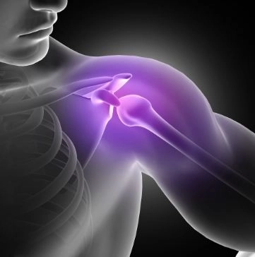
Sternoclavicular joint dislocation is considered a rare injury that many people may not have heard of. However, it is actually a condition that deserves serious attention, especially because it occurs in a sensitive area that connects the collarbone (clavicle) to the breastbone (sternum) in the center of the chest.At first, it may simply appear as mild pain or slight swelling after an injury or fall, but in reality, the severity of this condition can vary widely. In some cases, it may develop into a serious problem if it is not managed properly and promptly.The danger of sternoclavicular joint dislocation lies in its close proximity to vital structures within the chest, such as the major blood vessels, the trachea, and the esophagus. This makes accurate diagnosis and rapid medical intervention essential to prevent possible complications.Therefore, in my Dalily Medical guide, understanding this injury—its causes, symptoms, and treatment methods—is an important step for anyone interested in musculoskeletal health and overall body safety.
Is sternoclavicular joint dislocation dangerous?
In most cases of anterior dislocation, the injury is not considered highly dangerous and can often be managed with conservative treatment.
However, in the case of posterior dislocation, the condition can be serious, as it may lead to compression of:
- Major blood vessels
- The trachea (windpipe)
- The esophagus
For this reason, posterior dislocation is considered a medical emergency that requires prompt and careful intervention.
Does sternoclavicular joint dislocation always require surgery?
No, surgery is not always necessary.
In some cases, conservative treatment can be sufficient, which includes:
- Joint reduction (realignment)
- Immobilization
- Physical therapy
However, surgery may be required in the following situations:
- Posterior dislocation
- Unstable dislocation
- Chronic or recurrent cases
Can the joint return to its normal position on its own?
In some mild or partial cases, the condition may improve gradually over time without surgical intervention.
However, in complete dislocations, the joint usually does not return to its normal position spontaneously and requires medical treatment such as reduction, stabilization, or surgery depending on severity.
Does the pain last for a long time?
Pain is usually severe at the beginning of the injury, then gradually improves over a few weeks. However, mild pain or discomfort during shoulder movement may persist in some cases, especially if the injury is severe or if proper treatment and rehabilitation are not followed.
Does this injury affect arm movement?
Yes, it may temporarily affect arm movement due to pain and joint instability.
However, with proper treatment and adherence to physical therapy, shoulder function gradually improves, and most patients regain normal movement over time.
Types of sternoclavicular joint dislocation
Sternoclavicular joint dislocation (between the clavicle and sternum) can be classified based on severity and direction.
1) Based on severity
1. Partial dislocation (Subluxation)
- The joint is partially displaced, not fully separated
- Ligaments still hold part of the joint
- Moderate pain
- Limited shoulder movement
- Less severe than complete dislocation
2. Complete dislocation
- The clavicle fully separates from the sternum
- Clear displacement of the joint
- Severe pain with visible deformity
- Often requires urgent medical care
2) Based on direction
1. Anterior dislocation (forward)
- The clavicle moves forward
- Most common type
- Visible prominence at the upper chest
- Generally less dangerous than posterior dislocation
2. Posterior dislocation (backward)
- The clavicle moves backward toward the chest
- Most dangerous type because it may compress:
- Major blood vessels
- Trachea
- Esophagus
- May cause breathing or swallowing difficulties
3. Superior or inferior dislocation (rare)
- Very uncommon
- The clavicle moves upward or downward
- Usually caused by severe trauma or high-impact injuries
Causes of sternoclavicular joint dislocation
1) Direct trauma (most common)
- A strong blow to the shoulder or chest
- Car accidents (especially seatbelt-related injuries)
- Falling directly onto the shoulder or chest
2) Sports injuries
Contact sports such as:
- Football
- Wrestling
- Rugby
- Or injuries caused by falls or collisions during training
3) Indirect injuries
- Strong lateral force on the shoulder
- Sudden backward or forward movement of the shoulder
4) High-energy trauma
- Motorcycle accidents
- Falls from height
- Severe workplace injuries
5) Ligament weakness (less common)
- Joint hypermobility (loose ligaments)
- Certain conditions that weaken ligaments
- Previous injuries in the same joint
Symptoms of sternoclavicular joint dislocation
Symptoms vary depending on the type (anterior or posterior) and severity of the injury.
General symptoms
- Severe pain in the upper chest or at the base of the neck
- Swelling around the joint
- Visible deformity or prominence of the clavicle
- Difficulty moving the shoulder or lifting the arm
- Feeling of shoulder instability or weakness
Anterior dislocation (most common)
- Noticeable forward prominence of the clavicle
- Pain that worsens with movement or pressure
- Limited shoulder mobility
- Mild swelling or bruising in some cases
Posterior dislocation (most dangerous)
This type is considered an emergency because it may compress vital structures inside the chest.
Symptoms include:
- Severe chest pain or pain behind the sternum
- Difficulty breathing or a feeling of choking
- Difficulty swallowing
- Less obvious swelling compared to anterior dislocation
- Dizziness or palpitations in some cases due to vascular compression
Diagnosis of sternoclavicular joint dislocation
Diagnosis is very important because this injury may not be clearly visible on standard X-rays, especially in posterior dislocation cases.
1) Clinical examination
The doctor will:
- Take a detailed medical history (accident, fall, trauma)
- Examine the shoulder and upper chest
- Look for:
- Visible deformity or depression at the joint
- Pain on palpation
- Limited arm movement
- Compare both sides for asymmetry
2) X-ray imaging
- Usually the first imaging step
- Often not clear enough for this joint
- May appear normal even if injury exists
- Not reliable alone in many cases
3) CT scan (most important) ⭐
The most accurate diagnostic tool, showing:
- Direction of dislocation (anterior or posterior)
- Severity (partial or complete)
- Compression of blood vessels or vital organs
- Sometimes contrast is used for better vascular assessment
4) MRI scan
- Not essential in acute cases
- Useful for:
- Ligament evaluation
- Soft tissue and surrounding structure assessment
5) Special X-ray views
Such as the Serendipity view:
- Helps visualize the clavicle more clearly
- Used as an additional supportive tool
Medical (drug) treatment of sternoclavicular joint dislocation
Drug therapy is part of conservative treatment and aims to:
- Relieve pain
- Reduce inflammation and swelling
- Improve temporary mobility
⚠️ Important: Medications do not reposition the joint; they only relieve symptoms.
1) Pain relievers
Mild analgesics
- Paracetamol
Used for mild to moderate pain with limited anti-inflammatory effect
Non-steroidal anti-inflammatory drugs (NSAIDs)
Such as:
- Ibuprofen
- Diclofenac
- Naproxen
Benefits:
- Pain relief
- Reduction of inflammation and swelling
- Temporary improvement in movement
⚠️ Should be taken after food and not used long-term without medical advice
2) Muscle relaxants
Examples:
- Tizanidine
- Cyclobenzaprine
Benefits:
- Reduce muscle spasm
- Decrease pain related to muscle tension
- Improve comfort and sleep
3) Strong anti-inflammatory drugs (Corticosteroids)
- Not used routinely
- Only in selected cases under strict medical supervision
- Sometimes given as local injections
Benefits:
- Rapid reduction of severe inflammation
- Pain relief in difficult cases
⚠️ Must be used carefully due to potential side effects
4) Supportive treatment
- Ice packs during the first 48–72 hours
- Arm sling
- Limits movement
- Protects the joint
- Reduces pain
Important note
Drug treatment does not fix the dislocation itself. It is only used to control symptoms.
In some cases, especially:
- Posterior dislocation
- Unstable or complete dislocation
Patients may require reduction or surgical intervention.
⚠️ When does the condition become dangerous despite medication?
You should go to the emergency room immediately if any of the following symptoms appear:
- Shortness of breath
- Severe chest pain
- Difficulty swallowing
- Increasing or abnormal swelling
Surgical treatment of sternoclavicular joint dislocation (types and procedures)
Surgical treatment varies depending on several important factors, including:
- Type of dislocation (anterior or posterior)
- Severity (partial or complete)
- Presence of compression on blood vessels or vital organs
- Whether the injury is acute or chronic
Surgery is especially required in:
- Dangerous posterior dislocations
- Unstable dislocations
- Chronic cases that do not respond to conservative treatment
1) Closed reduction
What is it?
It is the process of realigning the joint without a large surgical incision.
Procedure:
- Performed under general anesthesia
- The doctor attempts to reposition the clavicle using controlled traction and manipulation
- External pressure or assistive tools may be used
Notes:
- Mostly used in recent injuries (within days)
- Lower success rate in posterior dislocations
- May require stabilization afterward
2) Post-reduction stabilization
Even after successful reduction, stabilization is needed to protect the joint.
Methods include:
- Arm sling
- Figure-of-8 brace
- In rare cases, temporary internal support devices
3) Open reduction surgery
What is it?
A direct surgical approach to expose the joint and restore alignment.
Steps:
- A small incision over the joint
- Exposure of the sternoclavicular joint
- Removal of any tissue blocking reduction
- Repositioning of the clavicle
- Surgical fixation of the joint
4) Ligament reconstruction ⭐ (most important)
Concept:
Instead of relying only on the joint alignment, the ligaments that stabilize it are rebuilt.
Method:
- Using a tendon graft from the patient’s own body (autograft)
- Or using strong synthetic ligaments
- The reconstruction is usually shaped in a figure-of-8 pattern between the clavicle and sternum
Goal:
- Long-term stability
- Prevention of recurrent dislocation
5) Fixation with screws or plates
Examples:
- Special screws
- Pins
- Small metal plates
Important notes:
- Not commonly used today
- Due to the proximity of vital structures
- Reserved for selected, carefully monitored cases
6) Medial clavicle resection
What is it?
Removal of a small portion of the inner end of the clavicle near the sternum.
When is it used?
- Chronic cases
- Persistent pain or arthritis
- Failure of other treatments
Goal:
- Reduce friction
- Relieve pain
- Improve mobility
7) Emergency posterior dislocation
In cases of compression symptoms:
- Immediate surgical intervention is required
- Sometimes involves vascular or thoracic surgeons
- Goal is to relieve pressure on vessels or the airway and stabilize the joint safely
After surgery
- Arm immobilization for 4–6 weeks
- Gradual physical therapy
- Avoid heavy lifting for a long period
- Follow-up imaging to ensure stability
⚠️ Important:
Surgery in this area is highly delicate due to its proximity to:
- The heart
- The lungs
- Major blood vessels
Tips for managing sternoclavicular joint dislocation
Proper management is essential to reduce pain, prevent complications, and speed recovery, especially because this joint is located near vital chest structures.
First: Immediate care after injury
1) Immobilize the arm
- Use a sling or supportive device
- Reduces movement and pain
- Prevents worsening of the injury
2) Apply ice packs
- 15–20 minutes per session
- Repeat every 2–3 hours for the first 48–72 hours
Benefits:
- Reduces swelling
- Relieves pain
- Decreases inflammation
3) Do NOT try to reposition the joint yourself
❌ Never attempt self-reduction at home
Because it may cause:
- Damage to blood vessels
- Worsening of the dislocation
- Serious complications
4) Go to the emergency room if severe symptoms appear
- Shortness of breath
- Severe chest pain
- Difficulty swallowing
- Abnormal swelling in the upper chest
Second: Early days management
1) Rest the shoulder
Avoid:
- Lifting the arm
- Carrying heavy objects
- Sudden movements
2) Take prescribed medications
- Painkillers and anti-inflammatory drugs as directed
- Do not stop treatment without medical advice
3) Proper sleeping position
- Sleep on your back
- Place a pillow under the injured arm
- Avoid sleeping on the affected side
Third: Recovery phase
1) Gradual physiotherapy
- Start with simple movements under supervision
- Progress gradually
Goals:
- Prevent stiffness
- Restore normal shoulder function
2) Avoid strenuous activity
Avoid:
- Heavy lifting
- Running or intense exercise
- Contact sports
3) Medical follow-up
- Regular check-ups
- CT scan in severe cases
- Ensure joint stability
Fourth: Long-term care
1) Strengthening exercises
- Light shoulder exercises
- Guided by a physiotherapist
2) Gradual return to activity
- Return to normal activity in stages
- Avoid sudden full load
3) Monitor warning signs
Seek medical advice if:
- Persistent or worsening pain
- Shoulder instability
- Visible deformity
- Numbness or weakness in the arm
⚠️ Common mistakes to avoid
- Attempting self-reduction
- Ignoring severe pain
- Returning to sports too early
- Neglecting serious symptoms like chest pain or breathing difficulty