

Many people complain of back or neck pain, often assuming it is nothing more than a simple muscle strain or fatigue from prolonged sitting. However, over time, they may discover that the underlying problem is far more serious. One condition that can be responsible for this type of pain is retrolisthesis, which occurs when a vertebra in the spine shifts backward abnormally. This displacement can place pressure on nearby nerves, leading to pain, numbness, and difficulties with movement.With today's lifestyle heavily dependent on sitting for long hours—whether at work, using computers, or scrolling on smartphones—and with the growing prevalence of excess body weight, an important question arises: Does prolonged sitting actually contribute to worsening symptoms? And can being overweight increase the severity of the condition or accelerate its progression?In this article from Dalili Medical, we will explore the relationship between prolonged sitting, excess weight, and retrolisthesis. We will also discuss practical ways to protect your spine, prevent further deterioration, and reduce pain through simple lifestyle changes.
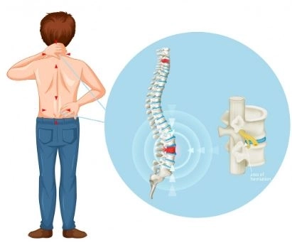
Retrolisthesis is a condition in which one vertebra moves backward, or slightly downward, relative to the vertebra directly beneath it. This displacement does not reach the level of a complete dislocation but can affect spinal stability and nerve function.
Not necessarily in all cases. Some cases are mild and stable and may cause few or no significant symptoms.
However, the condition becomes more concerning when it leads to:
Nerve compression
Severe or persistent pain
Weakness in the arms or legs
In most cases, the displaced vertebra does not return to its normal position spontaneously. However:
Symptoms can improve significantly with appropriate treatment.
Many patients can live a normal, active life if the condition remains stable and is properly managed.
In severe and very rare cases, retrolisthesis may result in significant nerve damage or serious neurological complications. However, complete paralysis is extremely uncommon and typically occurs only when there is severe, direct compression of the spinal cord.
Yes, the condition may progress if left untreated or if patients continue habits that place excessive stress on the spine, such as improper lifting techniques or neglecting core and back muscle strength.
However, with proper treatment and targeted strengthening exercises, the condition can often stabilize or improve considerably.
Yes. Sitting for extended periods can increase pain and place additional pressure on the spinal vertebrae.
For this reason, it is recommended to stand up, stretch, or move around every 30–45 minutes to reduce stress on the spine.
Yes. Being overweight increases the mechanical load on the spine, which may worsen pain and contribute to symptom progression.
Retrolisthesis is classified into several types based on the direction and degree of backward vertebral displacement relative to adjacent vertebrae. This classification helps physicians determine the severity of the condition and choose the most appropriate treatment plan.
In complete retrolisthesis, the affected vertebra shifts backward relative to both the vertebra above and the vertebra below it, making it noticeably displaced from its normal alignment.
In this type, the vertebra moves backward relative to only one adjacent vertebra—either the vertebra above or below. This is one of the most common forms of retrolisthesis.
This form involves progressive backward displacement of multiple vertebrae, creating a stair-step appearance on imaging studies. It is often associated with degenerative changes and age-related spinal wear and tear.
This affects the vertebrae of the neck and may cause:
Neck pain
Headaches
Numbness or weakness in the arms
This occurs in the mid-back region and is relatively uncommon because the thoracic spine is naturally more stable.
This is the most common type and affects the lower back. It may lead to:
Lower back pain
Pain radiating into the legs
Compression of the sciatic nerve (sciatica)
Retrolisthesis develops when factors affecting spinal stability allow a vertebra to shift backward. Common causes include:
As people age, the spinal discs lose flexibility and cushioning capacity, reducing spinal stability and increasing the risk of vertebral displacement.
The back and core muscles play a critical role in supporting the spine. Weakness in these muscles can reduce spinal stability and increase the likelihood of retrolisthesis.
Accidents, falls, or severe sports injuries can cause vertebrae to move out of their normal alignment.
Damage to the intervertebral discs can compromise spinal stability and contribute to backward vertebral slippage.
Osteoporosis weakens the vertebrae, making them more susceptible to movement, deformity, and instability.
Some individuals are born with structural abnormalities of the vertebrae or spinal joints that increase their risk of developing retrolisthesis.
Improper lifting techniques, repetitive bending, and chronic mechanical stress can place excessive pressure on the spine and contribute to instability.
Certain chronic conditions affecting the spinal joints and discs can gradually reduce spinal stability and lead to retrolisthesis over time.
The symptoms of retrolisthesis vary depending on the severity of the condition, the location of the affected vertebra, and whether nearby nerves are being compressed.
This is the most common symptom. The pain may be constant or triggered by movement and often worsens with prolonged standing, bending, or physical activity.
Patients may experience stiffness in the back or neck, particularly after prolonged sitting or upon waking in the morning. This can make movement more difficult and uncomfortable.
When the displaced vertebra compresses nearby nerves:
Pain may radiate into the legs if the lumbar spine is affected.
Pain may extend into the arms if the cervical spine is involved.
Compression of spinal nerves can cause numbness, tingling, or a "pins and needles" sensation in the legs, feet, hands, or arms.
Some patients may develop muscle weakness, which can make it difficult to walk long distances, climb stairs, or carry objects.
The muscles surrounding the spine may go into spasm as a protective response to stabilize the affected area.
In more advanced cases, nerve compression can interfere with coordination, balance, and normal walking patterns.
Symptoms often worsen during activities such as:
Lifting heavy objects
Prolonged sitting or standing
Sudden twisting or bending movements
If left untreated or improperly managed, retrolisthesis can lead to a range of complications, varying in severity depending on the degree of vertebral displacement and nerve involvement.
Over time, pain may become chronic and significantly affect daily life, leading to:
Sleep disturbances
Reduced mobility
Decreased productivity at work or school
Backward displacement of a vertebra can place pressure on spinal nerves, resulting in:
Numbness
Tingling sensations
Burning pain
Weakness in the limbs
When the lumbar spine is affected, compression of the sciatic nerve may occur, causing pain that radiates from the lower back down through the leg.
Persistent nerve compression can gradually weaken muscles in the arms or legs, depending on the location of the affected vertebra.
One of the more significant complications is spinal canal narrowing (spinal stenosis), which occurs when the space surrounding the spinal cord becomes reduced. This may cause:
Severe pain
Difficulty walking
Impaired balance
Abnormal movement between vertebrae can accelerate wear and tear of the spinal discs and facet joints, potentially worsening the condition over time.
Patients may experience limitations in performing everyday activities such as:
Bending
Sitting for extended periods
Lifting objects
Participating in sports or exercise
Advanced nerve compression may affect normal walking ability and overall balance, increasing the risk of falls.
In severe cases, substantial pressure on the nerves or spinal cord can result in marked weakness, reduced mobility, or partial loss of sensation.
This is one of the most serious complications and may indicate severe compression of the lower spinal nerves. It requires immediate medical evaluation and urgent treatment.
The diagnosis of retrolisthesis involves a combination of clinical evaluation and imaging studies to determine the presence, severity, and potential impact of the condition on surrounding nerves and spinal structures.
The diagnostic process begins with a detailed medical history. The physician may ask about:
The exact location of the pain
How long the symptoms have been present
The presence of numbness, tingling, or weakness in the limbs
Previous spinal injuries or trauma
Occupational and lifestyle factors, such as heavy lifting or prolonged sitting
A comprehensive physical examination is performed to assess spinal function and neurological status. This may include:
Evaluation of spinal range of motion
Muscle strength testing
Assessment of reflexes
Identification of pain during movement or palpation of the spine
X-rays are among the most important initial imaging tests because they can reveal:
Backward displacement of a vertebra
The degree of vertebral slippage
Signs of spinal degeneration or osteoarthritis
In some cases, the physician may request flexion-extension X-rays (taken while bending forward and backward) to assess spinal stability more accurately.
MRI is one of the most valuable advanced imaging techniques for evaluating:
Spinal nerves
Intervertebral discs
The spinal cord
The extent of nerve compression
An MRI is often recommended when symptoms include:
Numbness or tingling
Muscle weakness
Pain radiating into the arms or legs
A CT scan provides highly detailed images of the bony structures of the spine and is particularly useful in cases where surgical intervention is being considered.
In certain cases, physicians may order EMG and nerve conduction studies to evaluate:
The extent of nerve involvement
Potential impairment in nerve signal transmission
The severity of retrolisthesis is assessed based on several factors, including:
The degree of backward vertebral displacement
The presence and severity of nerve compression
The impact on mobility and daily function
The intensity of pain and associated neurological symptoms
Medication does not correct the vertebral displacement itself; however, it can help relieve pain, reduce inflammation, and manage associated symptoms such as muscle spasms and nerve pain. Treatment is typically tailored to the severity of the condition.
These medications are commonly used for mild to moderate pain.
Paracetamol (Acetaminophen)
Pain relief
Improved mobility and daily functioning
Note: Patients should follow recommended dosages to avoid potential liver-related side effects.
NSAIDs help reduce inflammation and pain associated with spinal irritation and nerve compression.
Ibuprofen
Diclofenac
Naproxen
Relief of back or neck pain
Reduction of inflammation and swelling
Improved movement and function
Possible side effects may include:
Stomach irritation
Heartburn
Increased blood pressure in some individuals
These medications should be used cautiously, particularly in patients with stomach, kidney, or cardiovascular conditions.
Muscle relaxants may be prescribed when muscle spasms or excessive muscle tension contribute to symptoms.
Cyclobenzaprine
Tizanidine
Reduction of muscle spasms
Relaxation of back muscles
Relief of pain associated with muscle tightness
Drowsiness
Dizziness
Reduced concentration
When retrolisthesis causes nerve compression resulting in numbness, tingling, or burning sensations, medications specifically targeting nerve pain may be recommended.
Gabapentin
Pregabalin
Relief of neuropathic pain
Reduction of numbness and tingling
These medications often require several days to several weeks before their full therapeutic effect is achieved.
Corticosteroids may be used in more severe cases to reduce inflammation around irritated nerves.
Prednisone
Epidural or targeted corticosteroid injections around the spine or affected nerves
Long-term corticosteroid use should be avoided unless closely supervised by a healthcare professional due to the risk of significant side effects.
Some patients benefit from topical therapies that help reduce localized pain and discomfort.
Anti-inflammatory gels
Pain-relief patches
Warming muscle creams
Relief of mild to moderate pain
Improved muscle comfort and relaxation
Surgical treatment for retrolisthesis is typically considered in advanced cases or when conservative treatments, such as medications and physical therapy, fail to provide adequate relief. The primary goals of surgery are to:
Stabilize the affected vertebrae
Relieve pressure on the nerves
Improve mobility and function
Reduce chronic pain
A physician may recommend surgical intervention in the following situations:
Severe, persistent pain that does not respond to conservative treatment
Weakness in the arms or legs
Significant compression of spinal nerves or the spinal cord
Difficulty walking
Loss of bladder or bowel control
Evident spinal instability
Spinal fusion is one of the most commonly performed procedures for treating retrolisthesis.
The surgeon permanently joins two or more vertebrae to prevent abnormal movement using:
Metal screws
Fixation rods or plates
Specialized spinal implants
Bone grafts (in some cases)
To stabilize unstable spinal segments and reduce pain caused by excessive vertebral movement.
Administration of general anesthesia
Surgical access through the neck or back
Realignment of the affected vertebra as close as possible to its normal position
Placement of screws, rods, or plates to secure the vertebrae
Closure of the surgical incision
This procedure is performed when retrolisthesis causes significant pressure on the spinal nerves.
Removal of a portion of bone
Removal of herniated or compressive disc material
Enlargement of the spinal canal
To relieve nerve compression and improve symptoms such as pain, numbness, and tingling.
A laminectomy involves removing part of the vertebra known as the lamina.
Creates additional space around the spinal cord and nerves
Reduces nerve compression
This procedure is frequently combined with spinal fusion to improve spinal stability and optimize outcomes.
This modern surgical technique uses small incisions rather than a traditional large surgical opening.
Less postoperative pain
Reduced blood loss
Faster recovery
Shorter hospital stay
The procedure utilizes specialized instruments and advanced camera-guided technology.
In selected patients, a damaged intervertebral disc may be replaced with an artificial disc.
To preserve spinal motion rather than permanently fusing the vertebrae.
Important: This option is not appropriate for all patients and is determined on an individual basis.
Larger incision
Direct visualization of spinal structures
Often used in more complex cases
Smaller incisions
Greater precision
Faster recovery and reduced postoperative discomfort
The recovery process may include:
A period of rest and activity modification
Physical therapy and rehabilitation
Gradual walking and mobility exercises
Avoidance of heavy lifting
Recovery time varies depending on the procedure performed and the patient's overall spinal condition, ranging from several weeks to several months.
As with any surgical procedure, complications may occur, including:
Infection
Bleeding
Nerve injury
Failure of spinal fusion (nonunion)
Persistence of some pain or symptoms
Despite these risks, many patients experience substantial symptom improvement following appropriate surgical treatment and postoperative care.
Therapeutic exercises are among the most effective non-surgical treatments for retrolisthesis because they help strengthen the muscles that support the spine and reduce stress on the vertebrae and nerves.
Strong abdominal muscles play an important role in supporting the spine and reducing pain.
Lie on your back.
Tighten your abdominal muscles by drawing them inward.
Hold for 5–10 seconds.
Repeat 10 times.
One of the most effective exercises for lower back stabilization.
Lie on your back with your knees bent.
Gently press your lower back toward the floor.
Tighten your abdominal and pelvic muscles.
Hold for 5 seconds, then relax.
Helps strengthen the lower back, gluteal muscles, and core.
Lie on your back with your knees bent.
Slowly raise your hips off the floor.
Keep your shoulders and feet firmly on the ground.
Hold for 5–10 seconds, then slowly lower your hips.
Improves spinal flexibility and reduces stiffness.
Position yourself on your hands and knees.
Arch your back upward like a cat.
Slowly lower and extend your back.
Repeat the movement in a smooth and controlled manner.
Stretching the hamstring muscles can reduce tension on the lower back.
Sit with one leg extended.
Lean forward gradually toward the extended leg.
Hold the stretch for 10–20 seconds.
Walking is one of the safest and most beneficial forms of exercise for many patients with retrolisthesis.
Improves circulation
Reduces stiffness
Naturally strengthens supporting muscles
It is generally recommended to start with 10–15 minutes per day and gradually increase the duration as tolerated.
Patients with retrolisthesis should generally avoid:
Heavy weightlifting
Sudden bending movements
High-impact running
Exercises that place excessive strain on the back
Rapid twisting or rotational movements of the torso
10 minutes of walking
10 pelvic tilts
10 abdominal contractions
5–10 bridge repetitions
Gentle stretching exercises
Daily self-care plays a crucial role in reducing pain and preventing disease progression, particularly in mild to moderate cases. The primary goal is to minimize stress on the spine while strengthening the muscles that support it.
Avoid sudden bending
Avoid rapid twisting motions
Do not lift heavy objects incorrectly
Keep your back straight while sitting
Use a chair with adequate lumbar support
Keep both feet flat on the floor
Avoid prolonged sitting and stand up every 30–45 minutes
Maintain good posture
Distribute body weight evenly on both feet
Walk with smooth, controlled steps
Avoid standing for long periods without movement
Sleeping on your back or side is generally recommended
Use a supportive pillow for proper neck alignment
Place a pillow between your knees when sleeping on your side
Avoid sleeping on your stomach, as it may increase spinal stress
Excess body weight places additional pressure on the spine.
Recommendations include:
Maintaining a healthy body weight
Following a balanced diet
Limiting excess sugar and unhealthy fats
Physical therapy is an essential component of treatment because it:
Strengthens the back and core muscles
Improves spinal flexibility
Reduces long-term pain
May help relax muscles and reduce muscle spasms.
May help reduce inflammation and alleviate pain.
Reduce prolonged sitting
Take regular movement breaks throughout the day
Engage in light walking regularly
Avoid excessive physical strain
A healthcare provider may recommend:
A lumbar support brace or back support belt
Important: Braces should not be used for extended periods without medical supervision.
Monitor symptoms regularly
Obtain imaging studies when necessary
Adjust the treatment plan according to symptom progression and clinical findings


