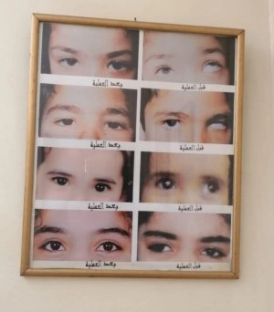
Some people notice a difference in the appearance of their eyes or their children’s eyes, where the distance between the inner corners of the eyes near the nose seems wider than normal, even though the eyes themselves are healthy and vision is unaffected. This condition is known as Telecanthus, a facial structural variation that may cause concern for some individuals, but in most cases it is not dangerous.The issue is not only cosmetic; it is also important to understand the underlying cause of this condition. Is it congenital from birth? Is it the result of an injury? Or is it related to abnormalities in the facial bones? This is where accurate diagnosis plays an important role in distinguishing between simple cases and those that may require medical follow-up or treatment.In this article from Dalili Medical, we will explore Telecanthus in detail: its causes, symptoms, degrees, possible complications, treatment options, and whether it can actually affect vision or not.
What is Telecanthus?
Telecanthus is a medical condition characterized by an increased distance between the inner corners of the eyes (medial canthi), while the pupils and eyeballs remain in their normal positions.
It is important to distinguish it from hypertelorism, where the actual distance between the eye sockets (orbits) is increased due to a skeletal bone abnormality.
Telecanthus is often associated with certain congenital facial or skull abnormalities, such as frontonasal dysplasia, which may also affect overall facial appearance.
❓ Is Telecanthus a serious condition?
In most cases, no ❌
Telecanthus is usually a cosmetic or structural variation that affects appearance only. However, in some cases, it may be a sign of an underlying congenital syndrome or facial bone development disorder.
❓ Does Telecanthus affect vision?
Generally, no
Vision and eye function remain completely normal because the condition affects only the position of the inner eye corners, not the eyeball itself or visual ability.
❓ What is the difference between Telecanthus and Hypertelorism?
- Telecanthus: Increased distance between the inner eye corners due to soft tissue or ligament involvement.
- Hypertelorism: Increased distance between the eye sockets due to bone structure enlargement.
❓ Can Telecanthus improve without treatment?
In mild cases, no treatment may be needed.
However, the condition usually does not improve on its own in moderate or severe cases and may remain unchanged unless medical intervention is performed.
❓ Is surgery safe?
Yes
Corrective surgery for Telecanthus is generally safe when performed by a specialist. However, like any surgical procedure, it may involve minor side effects such as:
- Temporary swelling around the eyes
- Mild bruising
- Slight asymmetry in rare cases
❓ Can Telecanthus return after surgery?
Rarely ❌
When the surgery is done properly and post-operative instructions are followed, results are usually stable and long-lasting.
❓ Can children be treated?
Yes
Doctors may treat children, but surgical correction is often delayed until facial bone growth is complete, except in severe cases that require early intervention.
❓ Is Telecanthus hereditary?
In some cases, yes
It may be linked to genetic factors or congenital syndromes, while in other cases it appears without a family history.
❓ Can it develop later in life?
Usually no ❌
Telecanthus is mainly congenital or trauma-related. However, it may become more noticeable after facial injuries or certain surgical procedures.
❓ Can Telecanthus be prevented?
Not always ⚠️
- Congenital cases: Cannot be prevented, as they occur during fetal development
- Acquired cases: May be reduced by preventing facial injuries and following safety measures
❓ Does Telecanthus cause pain or headaches?
No ❌
Telecanthus itself does not cause pain or headaches.
If symptoms exist, they are usually related to an underlying cause, such as trauma or associated bone abnormalities.
❓ Does Telecanthus affect psychological well-being?
Yes, in some cases ⚠️
Especially when the facial difference is noticeable, it may affect self-confidence or cause embarrassment. Psychological support and family acceptance play an important role, along with medical evaluation when needed.
❓ Do all cases of Telecanthus require a plastic surgeon?
No ❌
Mild cases do not require any treatment at all.
However, moderate or severe cases, or those associated with facial deformities, may require evaluation by a plastic surgeon or a maxillofacial surgeon.
❓ Can Telecanthus be corrected without surgery?
In most cases, no ❌
There are no exercises, creams, or non-surgical methods that can change the distance between the eyes.
Effective treatment is usually surgical or specialized cosmetic correction depending on the case.
❓ Can Telecanthus be a sign of a serious disease?
Sometimes, but rarely ⚠️
Telecanthus may appear as part of a congenital syndrome or a facial development disorder.
Therefore, doctors may request additional tests to rule out associated medical conditions.
Types of Telecanthus
Telecanthus is classified into several types depending on its cause and associated features:
1. Congenital (present at birth)
- Appears from birth
- Caused by abnormal development of midfacial bones or weak inner eye canthal ligaments
- May occur alone or as part of a genetic syndrome
- Most common type in children
2. Acquired Telecanthus
- Develops after birth at any age
- Causes include:
- Nasal or facial bone fractures
- Facial or eye surgery
- Severe facial trauma
- May appear suddenly or gradually after injury
3. Syndromic Telecanthus
- Part of a congenital syndrome or disorder
- Associated with chromosomal abnormalities or craniofacial syndromes
- Often accompanied by other facial changes (nose, eyelids, skull shape)
4. Pseudo-telecanthus (False Telecanthus)
- Actual bone structure is normal
- Appearance is due to external features such as:
- Excess skin near the inner eye corners
- Flat or wide nasal bridge
- Mainly a cosmetic appearance issue, not a structural defect
Causes of Telecanthus
Telecanthus is an increased distance between the inner corners of the eyes while the eyes themselves remain normal in size and function. The problem lies in positioning, not in the eyeball itself.
1. Congenital causes
- Abnormal facial bone development during fetal growth
- Genetic syndromes or chromosomal disorders
- Weak or underdeveloped inner eye ligaments
2. Trauma or injuries
- Fractures of the nasal or midface bones
- Direct facial injuries
- Previous surgeries affecting the central face or eye region
3. Syndromic conditions
- Craniofacial developmental disorders
- Genetic syndromes affecting facial structure
4. Bone abnormalities
- Deformities or imbalance in nasal or orbital bones
- Weak structural support in the midface
5. Rare causes
- Tumors in the midface region
- Severe infections affecting facial bones or tissues
Symptoms of Telecanthus
Symptoms are mainly cosmetic, as eye function is usually normal.
1. Increased distance between inner eye corners
- The main and most visible sign
- Eyes appear widely spaced even though eye size is normal
2. Facial asymmetry
- Midface may appear wider than normal
- Mild imbalance in facial proportions
3. Normal vision in most cases
- Visual acuity is unaffected
- Eye function remains normal
4. Associated features (depending on cause)
If part of a syndrome:
- Nasal shape differences
- Eyelid variations
- Mild craniofacial deformities
5. Psychological impact
- Feeling of being different in appearance
- Reduced self-confidence, especially in children and teenagers
Degrees of Telecanthus
Telecanthus is classified into degrees based on how much the distance between the inner eye corners increases compared to normal.
1. Mild (low grade)
- Slight increase in distance
- Barely noticeable except on close examination
- Mainly a cosmetic variation
- No effect on vision or eye function
2. Moderate grade
- More noticeable widening
- Mild facial imbalance
- Easily seen by others
- May be associated with nasal or eyelid changes
3. Severe grade
- Significant widening of inner eye distance
- Clear facial disproportion
- May be associated with midface bone deformities
- Often linked to congenital syndromes requiring medical evaluation
❓ Diagnosis of Telecanthus
The diagnosis of telecanthus relies on a series of careful steps to confirm the condition and rule out similar disorders.
1. Clinical examination
The doctor evaluates:
- The overall facial appearance
- The distance between the inner corners of the eyes
- The shape of the nose, eyelids, and facial bones to detect any additional deformities
2. Facial measurements
This is a key step in diagnosis and includes measuring:
- Intercanthal distance (distance between the inner eye corners)
- Interpupillary distance (distance between the pupils)
If the intercanthal distance is increased while eye position remains normal, telecanthus is likely.
3. Differentiating from similar conditions
- Telecanthus: Soft tissue or ligament issue affecting inner eye corners
- Hypertelorism: Increased distance between the eye sockets (bone-related condition)
4. Imaging tests
- CT scan of the face may be requested
- Helps evaluate nasal bones, skull structure, and detect deformities or trauma
- Especially useful in trauma or congenital syndrome cases
5. Evaluation of associated conditions
If a congenital syndrome is suspected:
- Full-body examination may be performed
- Genetic counseling may be recommended
⚠️ Complications of Telecanthus
In most cases, telecanthus is not a serious condition and mainly affects appearance. However, complications may occur depending on severity and underlying cause.
1. Cosmetic facial effects
- Noticeable widening between the eyes
- Facial asymmetry
- Possible dissatisfaction with appearance, especially in children and adolescents
2. Psychological and social effects
- Low self-confidence
- Feeling different from others
- Social withdrawal in some cases
3. Complications related to the underlying cause
If telecanthus is part of a syndrome:
- Nasal or facial bone deformities
- Skull development abnormalities
- Eyelid or orbital structural issues
4. Rare functional complications
In severe cases:
- Eyelid dysfunction (difficulty closing the eye completely)
- Soft tissue abnormalities affecting the eye region
5. Delayed diagnosis consequences
Ignoring the condition may delay detection of underlying syndromes or deformities, affecting treatment planning.
Medical (Drug) Treatment of Telecanthus
There is no direct drug treatment for telecanthus, as the condition is structural rather than infectious or inflammatory.
❌ Is there a medication for telecanthus?
No ❌
- No drugs can reduce the distance between the eyes
- Treatment is usually surgical or cosmetic correction
Medications used depending on the cause
1. In cases of injury or inflammation
- Anti-inflammatory drugs to reduce swelling
- Pain relievers
- Antibiotics if infection is present
These treat the cause, not the eye spacing itself.
2. In syndromic or developmental cases
- No direct medication for telecanthus
- Supportive treatments may be used depending on the patient’s condition
3. Eye-related symptoms
- Lubricating eye drops for dryness
- Anti-inflammatory eye drops if needed
4. Psychological support
- In rare cases, anti-anxiety or antidepressant medications may be prescribed under medical supervision
Surgical Treatment of Telecanthus
Surgical treatment aims to reduce the distance between the inner eye corners and improve facial symmetry, especially in moderate to severe cases or those associated with facial bone abnormalities.
❗ When is surgery needed?
- Moderate to severe telecanthus
- Noticeable cosmetic or psychological impact
- Associated craniofacial deformities
- After full facial bone growth (usually after childhood/adolescence)
Types of surgery
1. Medial canthoplasty
- Tightening or reconstructing inner eye ligaments
- Repositioning them to stronger bone structures
- Reduces inner eye distance without changing eye shape significantly
2. Osteotomy (bone correction surgery)
- Used when bone structure is the cause
- Midface or nasal bones are surgically adjusted
- Bones are repositioned inward and fixed with plates/screws
3. Soft tissue correction
- Removes or tightens excess skin or soft tissue
- Adjusts fat or eyelid structure
- Mainly used in pseudo-telecanthus
4. Craniofacial reconstructive surgery
- Used in severe or syndromic cases
- Multidisciplinary team approach
- Corrects bones, ligaments, and facial structure comprehensively
General surgical steps
- General anesthesia
- Small hidden incisions
- Correction of ligaments or bones
- Fixation with sutures or plates
- Cosmetic closure of skin
⚠️ Possible complications (rare)
- Temporary swelling or bruising
- Mild facial asymmetry
- Minor scarring
- Rare need for revision surgery
⏳ Recovery after Telecanthus surgery
First week
- Swelling and bruising around eyes
- Mild discomfort or tightness
- Cold compresses and pain relief
- Rest is essential
2–4 weeks
- Swelling decreases significantly
- Facial appearance improves
- Return to light daily activities
- Stitches removed within 5–10 days
4–8 weeks
- Noticeable cosmetic improvement
- Most bruising disappears
- Return to work or school
3–6 months
- Final results become fully visible
- Complete tissue healing
- Residual swelling resolves
Factors affecting recovery
- Type of surgery
- Patient age and health
- Post-operative care compliance
- Presence of complications
Prevention of Telecanthus
In most cases, telecanthus is congenital or trauma-related, so full prevention is not always possible.
1. Prenatal prevention (congenital cases)
- Genetic counseling if family history exists
- Regular pregnancy checkups and ultrasounds
- Avoid smoking, alcohol, and unsafe medications
- Proper nutrition, including folic acid intake
2. Prevention of acquired cases
- Protect face from injuries
- Use safety equipment during sports
- Early treatment of facial fractures
- Proper follow-up after facial surgery
3. Preventing worsening of the condition
- Early medical evaluation
- Regular follow-up with ophthalmology or maxillofacial specialists
- Timely intervention when needed