

Craniotomy surgery is one of the most important procedures in neurosurgery and is often performed to treat serious conditions such as brain tumors, bleeding, or head injuries. Although it is a major operation, advances in medical technology and modern techniques have made it much safer, helping patients regain their normal life after recovery.In this article, we will cover in detail everything you need to know: from the reasons for the surgery and its types, to the procedure itself, recovery time, and essential tips before and after the operation. The goal is that by the end of this article on Dely Medical, you fully understand the surgery and feel more informed and reassured.
What is a Craniotomy?
A craniotomy is a type of brain surgery where the surgeon temporarily removes a part of the skull (called a bone flap) to access the brain and treat the underlying issue. After the surgery, the bone is reattached using small plates and screws. Over time, the bone heals completely like any other broken bone and regains its natural strength.
Is Craniotomy Surgery Dangerous?
Although it is a major procedure, modern techniques have made craniotomy much safer. The success rate is high, especially when performed at the right time by an experienced neurosurgeon.
Does the Patient Feel Pain After Surgery?
Patients may experience mild to moderate pain at the surgical site, but it is usually temporary and easily controlled with pain medications.
Does the Surgery Affect Memory or Thinking?
In most cases, craniotomy does not cause any permanent effect on memory or cognition. Some patients may notice temporary difficulty with concentration or memory, which improves gradually over time.
Does Craniotomy Leave a Permanent Scar?
Typically, only a small surgical scar remains on the scalp, which usually fades over time and is covered by hair, making it barely noticeable.
Is the Surgery Performed Under General Anesthesia?
Yes, craniotomy is generally done under general anesthesia. In some specialized procedures, the patient may be partially awake during surgery to monitor speech or motor functions.
Can the Patient Feel the Skull Bone After Reattachment?
No, the bone is secured with small plates in a natural way, and the patient does not notice it in daily life.
Can the Skull Bone Move After Surgery?
This is very rare, as the bone is firmly fixed. By following the doctor’s instructions, the bone remains stable.
Does Surgery Affect the Shape of the Head?
Generally, the head’s shape does not change significantly. Mild swelling may appear initially but resolves gradually during recovery.
Can the Patient Sleep Normally After Surgery?
Yes, normal sleep is possible. It is recommended to choose a comfortable position and avoid pressure on the surgical site during the first few weeks.
Can the Patient Wash Their Hair After Surgery?
Yes, usually 7 to 14 days after surgery, with the doctor’s approval. Hair should be washed gently to protect the surgical site.
Can the Patient Exercise After Surgery?
Light walking: a few days after surgery
Light exercise: 4–6 weeks after surgery
Intense exercise: 2–3 months after surgery, with doctor’s permission
Can the Patient Travel After Craniotomy?
Yes, but it is recommended to wait 4–8 weeks and consult the doctor, especially before air travel.
Does the Surgery Affect Mental Health?
Some patients may experience temporary:
Anxiety
Mood swings
Mild depression
This is normal and improves over time with psychological support.
Can Seizures Occur After Surgery?
In some cases, seizures may occur. Doctors often prescribe temporary preventive medications.
Is Physical Therapy Needed After Surgery?
Yes, in some cases, especially if there is weakness or balance issues. Physical therapy helps restore strength and mobility.
Can the Surgery Be Repeated?
Yes, if needed, for example:
Tumor recurrence
A new brain problem requiring surgery
Does the Surgery Affect Life Expectancy?
In most cases, craniotomy helps save lives and improves life expectancy, especially when serious conditions are treated early.
Can the Patient Use Phone or Computer After Surgery?
Yes, but initially for short periods to avoid eye or head strain.
Can the Patient Fully Return to Normal Life?
Yes, many patients return to their normal life completely after full recovery and regain most daily functions.
Craniotomy involves temporarily opening a part of the skull to access the brain, then replacing the bone after surgery. It is used to treat serious brain conditions.
Common Reasons Include:
Brain Tumors
Removing benign or malignant brain tumors
Taking a biopsy to determine tumor type
Reducing pressure from the tumor on the brain
Goal: Protect brain function and reduce symptoms like headaches and seizures
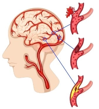
Brain Hemorrhage
Caused by:
Severe head injuries
Blood vessel rupture
Hemorrhagic stroke
Goal: Remove blood accumulation, reduce pressure on brain tissue, prevent permanent damage
Severe Head Injuries
From accidents or falls:
Brain swelling
Brain contusions
Skull fractures
Role of Surgery: Relieve pressure and prevent serious complications
Brain Aneurysms
Weakness in a blood vessel wall causing ballooning
Goal: Repair the aneurysm and prevent rupture or serious bleeding
Clots or High Intracranial Pressure
Caused by:
Severe swelling
Large clot
Fluid accumulation in the brain
Goal: Relieve pressure and save the patient’s life
Certain Neurological Disorders
Severe epilepsy not responding to medication
Serious infections like brain abscess
Vascular malformations
Foreign Object Removal
In cases like:
Gunshot injuries
Objects entering the skull
Role of Surgery: Safely remove foreign objects and protect the brain
Craniotomy varies depending on the skull opening location and surgical goal. Each type allows the surgeon to access a specific brain area.
Frontal Craniotomy
Location: Front of the skull (forehead)
Use: Frontal lobe tumors, front brain injuries, some epilepsy cases
Advantage: Access to thinking and behavior areas
Temporal Craniotomy
Location: Side of the head above the ear
Use: Temporal lobe tumors, treatment-resistant epilepsy, some memory disorders
Parietal Craniotomy
Location: Upper side of the skull
Use: Parietal lobe tumors, bleeding, swelling in this area
Occipital Craniotomy
Location: Back of the head
Use: Occipital lobe tumors, vision problems due to brain injuries
Suboccipital Craniotomy
Location: Lower back of the skull
Use: Cerebellar tumors, brainstem tumors, balance issues
Pterional Craniotomy
Location: Temporal region near the eye
Use: Brain aneurysms, skull base tumors
Note: One of the most common types
Keyhole Craniotomy
Method: Very small opening
Advantages: Smaller incision, less pain, faster recovery, fewer complications
Use: Small tumors or certain bleeding cases
Procedure: Opening a larger portion of the skull
Uses:
Large tumors
Severe injuries
Extensive bleeding
Purpose: To relieve high intracranial pressure caused by:
Brain swelling
Severe bleeding
Serious head injuries
Note: In some cases, the bone flap may be left out temporarily to reduce pressure.
Craniotomy: The skull bone is replaced after surgery.
Craniectomy: The bone is not immediately replaced, usually to relieve intracranial pressure.
The method depends on the location, size, and type of brain problem. The main goal is to reach the affected area safely while minimizing damage to healthy tissue.
Procedure Steps:
General anesthesia is administered.
An incision is made on the scalp in the forehead area.
Skin and tissues are gently moved aside.
A surgical drill opens a section of the skull.
The bone flap is lifted.
The frontal lobe is accessed to treat the problem (tumor, bleeding, etc.).
The bone is replaced and secured.
The scalp is closed with sutures.
Procedure Steps:
An incision is made above the ear or on the side of the head.
The skull in the temporal region is exposed.
The bone is opened with precise tools.
The temporal lobe is accessed.
The tumor is removed or bleeding repaired.
The bone is replaced and the incision closed.
Procedure Steps:
The patient is placed on the stomach or side.
An incision is made at the back of the head.
The posterior skull is exposed.
Part of the bone is removed to reach the cerebellum or brainstem.
The tumor or problem is treated.
The bone is replaced and the incision closed.
Procedure Steps:
Incision near the hairline in the temporal region.
Muscles and tissues are moved aside.
A small portion of the skull is opened.
The skull base is accessed.
Aneurysms are repaired or tumors removed.
The bone is replaced and the incision closed.
Procedure Steps:
A very small incision (2–3 cm) is made.
A small opening in the skull is created.
Precise tools or an endoscope are inserted.
The problem is treated without a large opening.
The incision is closed with sutures.
Advantages: Smaller wound, less pain, faster recovery.
Procedure Steps:
A relatively large incision is made.
Part of the skull is removed to reduce pressure on the brain.
In some cases, the bone is not immediately replaced.
The skin may be temporarily closed only.
General anesthesia
Head stabilization to prevent movement during surgery
Use of a surgical microscope for precision
Monitoring brain function during surgery
Bone replacement and fixation with small plates
Wound closure and scalp suturing
Diagnosing the need for craniotomy is very precise. Doctors rely on clinical exams and imaging to determine if a brain problem requires surgery. According to protocols from specialized centers like Mayo Clinic and Cleveland Clinic, the process involves:
Neurological Clinical Exam
Muscle strength in arms and legs
Reflexes
Balance and gait
Vision
Speech
Memory and concentration
Goal: Identify any malfunction in a specific brain region
Magnetic Resonance Imaging (MRI)
Detects brain tumors, swelling, infections, and structural abnormalities
Provides highly detailed images of brain tissue
Computed Tomography (CT Scan)
Especially useful in emergencies
Diagnoses intracranial bleeding, skull fractures, head injuries, clots
Fast and suitable for emergency cases
Angiography
Examines brain blood vessels
Detects aneurysms, arteriovenous malformations, and blockages
Electroencephalography (EEG)
Used in epilepsy or seizure cases
Determines abnormal brain activity source
Blood Tests
Evaluate overall health
Detect infections
Assess body’s ability to tolerate surgery
Brain Biopsy (in some cases)
A small tissue sample is taken
Determines tumor type accurately
Helps plan proper treatment
Craniotomy is a major brain surgery. While it can save lives or treat serious conditions, it may have some risks. These vary depending on the reason for surgery, patient age, health status, and surgical team skill.
Pain: Head or surgical site pain, usually resolves in days/weeks, managed with painkillers
Swelling and bruising: Appears on scalp or face, usually resolves in 1–2 weeks
Headache: Common after brain surgery, usually temporary
Temporary weakness or paralysis: Arm or leg weakness, usually improves with therapy
Speech problems: Temporary difficulty speaking, especially if surgery is near language areas
Memory or concentration issues: Usually temporary, improves over time
Loss of balance: Common in cerebellar surgeries
Bleeding: May occur during or after surgery, rarely requires further intervention
Infection: Rare, can occur at the wound or inside the brain (symptoms: fever, redness, swelling, discharge)
Seizures: May occur post-surgery; preventive medications are given
Brain swelling: Can increase intracranial pressure, managed medically or surgically
Cerebrospinal fluid leak: May require medical attention
Stroke
Permanent brain damage
Coma or life-threatening complications
Older age
Chronic diseases (diabetes, heart disease)
Size and location of brain problem
Length of surgery
Delayed treatment
Recovery depends on the reason for surgery, type of surgery, patient age, and health. Generally, recovery passes through stages:
Hospital Recovery
Duration: 3–7 days, sometimes 10–14 days for complex cases
Monitoring brain function and consciousness
Administering painkillers and medications
Wound care
Gradual movement
First 2–4 Weeks Post-Discharge
Fatigue, mild headache
Rest, avoid exertion
Gradual wound healing
Light activities like indoor walking
Intermediate Recovery (4–8 Weeks)
Gradual improvement in energy
Return to some daily activities
Pain reduction
Brain begins to recover and adapt
Some may need physical therapy
Full Recovery (2–6 Months)
2–3 months for simple cases, up to 3–6 months for complex cases
Resume work and normal activities
Restore most daily functions
Reason for surgery (tumor, bleeding, injury)
Surgery size
Patient age and general health
Adherence to doctor’s instructions
Office work: 4–8 weeks
Driving: 4–6 weeks (with doctor approval)
Heavy exertion: 2–3 months
Improved consciousness
Gradual disappearance of headaches
Wound healing
Improved mobility and speech
Before Surgery:
Complete medical tests (MRI, CT, blood tests, ECG)
Inform doctor of all medications (blood thinners, diabetes or blood pressure drugs)
Stop eating 6–8 hours and drinking 4–6 hours before surgery
Shower and wash hair before surgery
Maintain mental calmness and trust the medical team
Stop smoking
After Surgery:
Get enough rest
Keep wound clean and dry, avoid touching it
Take medications as prescribed (painkillers, antibiotics, anti-seizure meds)
Avoid heavy activities (lifting, bending, vigorous exercises) for 4–8 weeks
Gradual walking to improve circulation and speed recovery
Healthy nutrition: protein, vegetables, fruits, and enough water


