

In modern medicine, X-rays are considered one of the essential diagnostic tools used to detect health problems in various parts of the body, including the teeth, bones, and internal organs. Among the advanced imaging technologies is the Focused Beam X-ray, which allows doctors to obtain highly accurate and reliable images of specific targeted areas.In this article from Dalili Medical, we’ll explore what Focused Beam X-ray is, how it works, its benefits, applications in both dentistry and general medicine, and how it compares to other types of X-ray imaging.
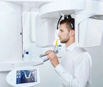
A Focused Beam X-ray is a specialized type of dental X-ray that directs a narrow, precisely aimed beam at a specific area—such as a particular tooth or segment of the jaw. Unlike traditional X-rays that use a broader, unfocused beam, this method enhances image clarity and detail while minimizing the exposure of other tissues to radiation.
The process follows the same principles as conventional X-ray techniques but with enhanced precision:
Patient Preparation
The clinician positions the patient carefully, targeting the region of interest—such as a particular tooth or root area.
Focused X-ray Beam Activation
The device emits a tight beam of X-rays directly at the selected target, using a digital sensor or film to capture the image.
X-ray Exposure
The focused beam passes through the targeted structures, with minimal scattering or impact on surrounding areas.
Image Capture and Analysis
A sensor records the X-ray data, which is then reviewed on a screen or printed film to detect specific dental or structural issues.
High-Resolution Imaging: Offers crisp, detailed images ideal for identifying subtle issues like small cavities or early-stage root infections.
Lower Overall Radiation Exposure: By confining the beam to a small area, it significantly reduces unnecessary radiation to adjacent tissues.
Reduced Image Artifact: The targeted beam helps minimize image distortion and enhances diagnostic accuracy.
Rapid Diagnosis: Clinicians can quickly pinpoint and address dental or anatomical issues with minimal wait time.
This focused imaging method is highly useful in several dental scenarios:
| Common Use | Description |
|---|---|
| Cavity Detection | Identifies early-stage decay in areas that are hard to visualize, like between molars. |
| Root Pathology | Detects infections, abscesses, or inflammation around tooth roots. |
| Implant Planning | Offers precise visualization of bone structure and density crucial for implant placement. |
| Orthodontic Monitoring | Helps assess root alignment and jaw structure during orthodontic treatment. |
| Bone Health Evaluation | Identifies areas of bone loss or structural weakness around the teeth. |
Beyond dentistry, Focused Beam X-rays are valuable in medical settings too:
Fracture Diagnosis: Particularly in fine or small bones where detail is crucial.
Detecting Small Lesions or Infections: Ideal for examining localized areas in soft tissues or organs.
Surgical Planning: Helps map minute anatomical details before surgeries.
Precision: Pinpoints specific areas for enhanced diagnostic clarity.
Safety: Targets only the needed area, reducing excess radiation exposure.
Sharper Imagery: Narrow beam decreases scatter and increases diagnostic reliability.
Patient-Friendly: Typically quick and comfortable for the patient, with minimal setup time.
Radiation Exposure: Though reduced, exposure still occurs—especially with repeated use.
Not Ideal for Wide-Area Diagnosis: If broader oral or anatomical challenges exist, more comprehensive imaging (like full-mouth or panoramic X-rays) may be necessary.
Expert Interpretation Required: Accurate results depend on both correct beam placement and skilled reading of the resulting images.
In both modern dentistry and medical diagnostics, Focused Beam X-rays are a cutting-edge tool offering high precision, reduced radiation, and exceptional imaging clarity. Whether detecting dental decay, guiding implant therapy, or planning delicate procedures, this technology ensures better, more targeted care. For challenges requiring pinpoint imaging with minimal patient exposure, Focused Beam X-rays are an outstanding solution.


