
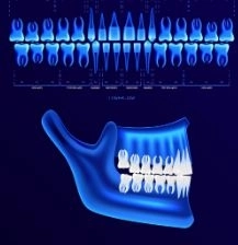
Cone Beam CT (CBCT) imaging is considered one of the latest technologies in the field of dentistry, which has revolutionized the way many dental conditions are diagnosed and treated. This technology represents a major advancement compared to traditional techniques such as conventional X-rays, as it provides highly accurate three-dimensional images of the teeth and jaws. In Dalily Medical, we will learn about Cone Beam CT, how it works, its benefits, its importance in diagnosing and treating dental and jaw problems, as well as a comparison with other imaging techniques.
Cone Beam CT (CBCT) is a type of computed tomography used to image the teeth and jaws in three dimensions. The device captures a series of X-ray images in a spiral (semi-circular) motion, which are then processed to create a 3D image of the targeted area. This technique differs from conventional CT scans in that it uses a cone-shaped X-ray beam, allowing for highly precise imaging. This makes it ideal for diagnosing problems that are difficult to detect using other imaging methods.
Cone Beam CT works by directing a cone-shaped X-ray beam at the target area in the mouth and jaws. The X-rays are captured by a special detector while the machine rotates around the patient, taking images from different angles. These images are then processed instantly by a computer to produce a detailed 3D image of structures such as teeth, bones, nerves, and surrounding blood vessels.
1. Preparation:
The patient stands or sits in a specific position to properly align the jaw and mouth with the scanner.
2. Scanning:
The X-ray beam is directed at the target area while the machine rotates around the patient, capturing multiple images.
3. Image processing:
The collected images are converted into a three-dimensional image by computer software.
4. Analysis:
The dentist analyzes the detailed 3D images to make an accurate diagnosis and determine the best treatment plan.
CBCT provides highly detailed 3D images that allow dentists to identify dental and jaw problems with exceptional precision. Unlike traditional X-rays, which produce 2D images that may miss important details, CBCT offers a complete and realistic view of issues such as deep caries, jaw deformities, and impacted teeth.
With the accurate diagnosis provided by CBCT, dentists can make better-informed treatment decisions. In some cases, this technology helps avoid unnecessary surgical interventions, reducing pain and treatment time.
This technology enables 3D evaluation of the targeted area, making it ideal for complex cases such as dental implants or jaw abnormalities. It helps determine the optimal implant position and assess fractures or misalignments with high accuracy.
One of the key advantages of CBCT is its lower radiation dose compared to conventional CT scans, making it safer for patients, especially children and pregnant women.
Assessing bone and surrounding tissues is essential for treatment planning. CBCT provides detailed information about bone structure, which is particularly useful for procedures such as dental implant planning.
Cone Beam CT is considered one of the essential tools in dental implant planning. By accurately imaging the bone surrounding the target area, the dentist can determine the optimal implant position with high precision. In addition, CBCT helps assess bone thickness and detect any structural issues that may affect implantation.
CBCT is used to diagnose jaw problems and deformities such as misalignment or structural abnormalities. It is also used in cases that require orthodontic treatment or jaw surgery.
In cases that cannot be easily diagnosed using traditional X-rays—such as hidden cavities or subgingival decay—CBCT is an ideal tool for detecting such issues. It is also used to monitor the progress of orthodontic treatment.
This imaging technique helps detect cysts or tumors in the mouth and jaw, especially those that are not visible or difficult to identify using conventional examinations.
CBCT is also used to monitor treatment success after dental surgery or implant placement. The dentist can evaluate bone healing and verify implant positioning within the bone.
One of the main differences between CBCT and conventional X-rays is the ability to produce three-dimensional images. This feature allows dentists to obtain more accurate and realistic views of complex dental structures.
As previously mentioned, CBCT uses a lower radiation dose compared to conventional CT scans, making it safer for patients.
CBCT is faster to perform than traditional X-rays or CT scans. It provides nearly immediate results, which helps speed up treatment decisions.
One of the main challenges of CBCT technology is the high cost of the equipment. This may limit its availability in some dental clinics and centers.
Due to the complexity of the technology and the use of 3D imaging, dentists and technicians require specialized training to ensure accurate results.
Despite its advantages, CBCT is not suitable for all cases. Some dental conditions may still require a combination of CBCT and traditional X-rays for a complete diagnosis.