

Bitewing X-ray is one of the most widely used techniques in dentistry for detecting cavities and gum diseases effectively. This imaging technique is essential for the early detection of many dental health issues, especially those that are difficult to spot through a visual examination alone. In this article, we will guide you through a detailed explanation of the importance of this X-ray, how it works, and the benefits it offers to patients.
Bitewing X-ray is a type of radiographic imaging primarily used to detect cavities between teeth and assess the health of the supporting bone structures of the teeth. This X-ray is performed by placing a small imaging device inside the patient's mouth, and the patient is asked to "bite" down on the device to allow a clear image of the back teeth to be captured.
Bitewing X-rays are distinctive because they show teeth in their natural alignment, making the images extracted from these X-rays valuable for detecting issues that may not be visible during a clinical examination.
At the beginning of the procedure, the dentist places a small imaging device or film inside the patient’s mouth. The patient is then asked to bite down on the device in a specific way. This position helps capture a clear image of both the upper and lower teeth simultaneously.
The X-ray is directed through the patient's mouth to capture an image of the teeth. It is essential for the angle between the X-rays and the teeth to be precise, as incorrect angles can result in blurry or ineffective images. Typically, a 10-15 degree angle is used to capture the image correctly.
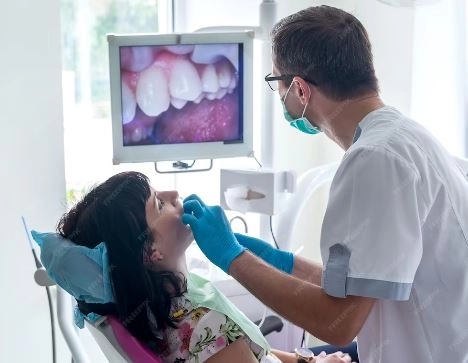
Today, most dental X-rays use digital technology instead of traditional films, allowing the dentist to receive a clear image instantly on the computer. Additionally, this technology reduces the amount of radiation exposure for the patient.
One of the main uses of bitewing X-rays is detecting cavities that develop between teeth, which may not be visible during a regular visual examination. This type of X-ray allows the dentist to assess the extent of the cavity, enabling early treatment before it worsens and leads to pain or tooth loss.
Bitewing X-rays are also used to evaluate the health of the bones that support the teeth. These X-rays can help identify issues such as bone loss due to gum disease. The X-ray can reveal changes in bone density, which may indicate inflammation or other health problems.
Bitewing X-rays are used to assess the condition of dental fillings and determine whether there is decay around them. They also help examine the condition of teeth after orthodontic treatment to ensure they are in good position and no cavities are forming around the wires or brackets.
For children, these X-rays can be used to evaluate tooth growth and detect potential problems early on. For example, if a child is developing cavities between teeth, the X-ray can identify the issue before it becomes noticeable to the parent or even the dentist.
Bitewing X-rays are one of the most effective tools for detecting cavities between teeth. They can reveal small or hidden cavities that are not visible to the naked eye, allowing dentists to address the problem early on.
These X-rays also provide accurate information about the condition of the bone surrounding the teeth, helping detect gum diseases such as gingivitis or bone loss before they escalate into major problems.
With digital X-ray technology, bitewing X-rays have become more precise and efficient. Digital images do not require processing like traditional films, meaning the dentist receives results faster and does not need to repeat the examination.
The amount of radiation exposure during a bitewing X-ray is significantly lower compared to other types of radiographic imaging, such as panoramic X-rays or traditional X-rays.
Despite the numerous benefits of bitewing X-rays, there are some risks associated with their use, especially if overused or performed in inappropriate circumstances.
While the radiation from bitewing X-rays is lower compared to other types, repeated exposure to X-rays can have negative effects on the body, particularly for children and pregnant women.
If the X-ray is not positioned properly or if the angles are not accurate, the images may be unclear, which could lead to an incorrect diagnosis or the need for a follow-up examination.
Bitewing X-rays typically focus on the back teeth and may not provide a good assessment of the front teeth. As a result, the dentist may need to use other types of X-rays to get a comprehensive evaluation of the teeth.
In recent years, digital X-rays have become the preferred option in most dental clinics. Digital X-rays offer several advantages over traditional films, including:
Faster Images: The images are immediately displayed on a computer, saving both time for the patient and the dentist.
Higher Quality: Digital images provide greater detail and clarity, helping the dentist to make more accurate diagnoses.
Reduced Radiation: Digital X-rays reduce the amount of radiation the patient is exposed to by up to 80%.
No, bitewing X-rays cannot replace a traditional clinical examination. While they help identify many dental health issues, the clinical exam is still essential for determining overall oral health. It is always recommended to combine both the clinical exam and bitewing X-rays for the best assessment of oral health.
Bitewing X-rays are a vital tool in dentistry for the early detection of cavities and other dental health issues. They help ensure the health of your teeth and gums, offering a precise and effective way to diagnose and treat dental problems early, saving time, money, and discomfort in the long run. Ultimately, oral health remains a top priority for overall well-being, and utilizing this advanced technology can contribute to preventing many dental health problems.


