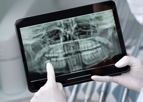
In the world of dentistry and oral and maxillofacial surgery, the temporomandibular joint (TMJ) is considered one of the most complex joints in the human body. It connects the lower jaw to the skull and enables essential functions such as chewing, speaking, and swallowing. When this joint develops problems, it can cause distressing symptoms such as pain, clicking sounds, and difficulty opening or closing the mouth. Here at Dalili Medical, TMJ imaging serves as an essential diagnostic tool, helping doctors accurately assess the condition and develop an effective treatment plan—especially with the significant advancements in imaging technology such as CBCT and MRI.
What is the temporomandibular joint (TMJ) and what is its function?
The temporomandibular joint (TMJ) is a paired joint located just in front of each ear. It connects the lower jaw (mandible) to the skull and is one of the most complex joints in the human body.
It consists of:
- The condyle of the mandible (the head of the lower jaw bone)
- The temporal bone socket in the skull
- An articular disc (a cartilage pad) that acts as a shock absorber between the bones
Functions of the TMJ:
- Opening and closing the mouth
- Moving the jaw forward, backward, and side-to-side
- Assisting in chewing (mastication)
- Supporting speech and swallowing
Why is TMJ imaging performed?
Doctors request TMJ imaging in the following cases:
- Chronic jaw or ear pain
- Difficulty or limitation in opening the mouth
- Clicking or popping sounds in the joint
- Swelling or inflammation around the TMJ
- Jaw injuries or fractures
- Pre-surgical evaluation
- Joint disorders such as disc displacement or cartilage degeneration
Types of TMJ imaging
1. Conventional X-ray
- Mainly shows bone structures
- Useful for detecting fractures or major bone abnormalities
- Does not clearly show soft tissues or the joint disc
2. Panoramic X-ray
- Provides a broad overview of the teeth, jaws, and TMJ
- Quick and simple to perform
- Less detailed compared to advanced imaging techniques
3. Cone Beam CT (CBCT)
- Provides high-resolution 3D images of the jaw and TMJ
- Excellent for detecting bone changes, fractures, and joint degeneration
- Uses lower radiation dose compared to traditional medical CT
4. Magnetic Resonance Imaging (MRI)
- Best for visualizing soft tissues
- Shows the joint disc, ligaments, and surrounding muscles
- Does not use radiation, but is more expensive
5. Ultrasound
- Safe and fast method
- Useful for detecting inflammation or fluid accumulation
- Limited in showing deep internal joint structures
Steps of TMJ imaging examination
1. Initial evaluation
- Reviewing symptoms and medical history
- Choosing the appropriate imaging technique
2. Preparation
- Removing jewelry or metal objects
- In some cases, the patient may be asked to open and close the mouth during imaging
3. Imaging procedure
- Depends on the selected method (X-ray, CBCT, MRI, etc.)
- The patient may sit or lie still while the head is stabilized
4. Results analysis
- The doctor examines the images to identify the problem
- A treatment plan is then developed
Benefits of TMJ imaging
- Accurate diagnosis of bone and soft tissue problems
- Assessment of bone erosion or cartilage damage
- Precise planning for jaw surgery
- Monitoring response to treatment
Risks and precautions
Although most imaging techniques are considered safe, there are some important precautions to keep in mind:
- Avoid unnecessary exposure to radiation
- Extra caution in pregnant women, especially when using X-ray–based imaging
- Check for contrast material allergies if contrast agents are used in MRI or CT scans
Cost of TMJ (temporomandibular joint) imaging
The cost varies depending on the type of examination and the medical center:
- X-ray: the lowest cost option
- CBCT (Cone Beam CT): moderate cost
- MRI: the highest cost due to advanced soft tissue imaging
Recent advances in TMJ imaging
Modern technology has significantly improved the accuracy and efficiency of TMJ diagnosis:
- Integration of artificial intelligence (AI) in image analysis for faster and more precise diagnosis
- Development of low-dose radiation imaging techniques to improve patient safety
- Introduction of portable imaging devices for TMJ evaluation in smaller clinics
Conclusion
Imaging of the temporomandibular joint (TMJ) is not just a diagnostic tool—it is a crucial step in determining the appropriate treatment and preventing complications.
With continuous technological advancements, it is now possible to obtain highly accurate images with minimal radiation exposure and in a short time, helping both doctors and patients achieve better treatment outcomes.