

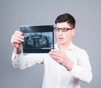
With the rapid advancements in the field of dentistry, 3D dental imaging has emerged as one of the most important advanced diagnostic technologies, revolutionizing the accuracy of examinations and treatment planning. This technique relies on Cone Beam Computed Tomography (CBCT) to produce high-resolution images that reveal precise details of the mouth and jaws from all angles. It enables dentists to diagnose complex cases such as root problems, bone loss, or optimal planning for dental implants. Thanks to its speed and precision, 3D dental imaging has become an essential tool for anyone seeking accurate diagnosis and successful treatment in Dalili Medical modern dentistry.
What is 3D Dental Imaging?
3D dental imaging is an advanced diagnostic technique that uses specialized devices such as Cone Beam Computed Tomography (CBCT) to produce high-resolution, three-dimensional images of the oral and jaw structures.
These images are reconstructed using specialized computer software to provide a comprehensive view of the teeth and surrounding bone, giving the dentist the ability to study fine details from all angles.
Comprehensive and Accurate View
Reveals the finest details in bones, teeth, and root canals.
Reduced Margin of Error
Helps plan treatments accurately and avoid surprises during procedures.
Greater Safety
CBCT devices often emit lower radiation doses compared to traditional medical CT scans.
Fast Scanning
The scanning process takes only a few seconds, with immediate results available.
Dental Implants
Determines the ideal implant placement.
Measures bone density and height.
Avoids injury to nerves or the sinuses.
Endodontics (Root Canal Treatment)
Detects complex or additional root canals.
Assesses the success of root canal therapy.
Identifies fine root fractures.
Oral and Maxillofacial Surgery
Studies fracture or deformity cases.
Plans orthognathic (jaw correction) surgeries.
Periodontal and Bone Diseases
Measures bone loss caused by gum disease.
Monitors the progression of the condition after treatment.
Tumor and Cyst Detection
Identifies the location and size of bone tumors or cysts.
Evaluates their impact on surrounding structures.
Initial Evaluation
Review the patient's medical history and ensure the scan is appropriate.
Preparation
Remove jewelry or removable metallic restorations.
Scanning Procedure
The patient is asked to stand or sit inside the CBCT machine, remaining still for a few seconds.
Image Processing
Images are transferred to a computer for 3D reconstruction and display.
Result Analysis
The dentist interprets the images and develops the treatment plan accordingly.
More precise treatment planning.
Reduced surgical risks.
Early detection of hidden problems.
Improved treatment success rates.
Although 3D dental imaging is considered safe, some points should be kept in mind:
Radiation exposure (although minimal) should be limited to necessary cases.
Avoid its use for pregnant women unless absolutely necessary.
Inform your dentist of any special medical conditions.
Use modern devices with low radiation doses.
Have the scan done in specialized centers.
Follow your dentist’s instructions during the scan.
Recent advancements aim to integrate artificial intelligence with 3D dental imaging to provide accurate diagnoses in seconds, while further reducing radiation exposure. Portable devices are also being developed, making scans possible anywhere.
Advanced 3D dental imaging has revolutionized diagnosis and treatment planning, offering dentists a clear and precise view, and providing patients with greater comfort and safety. With ongoing developments in imaging technology, this technique is expected to become even more widespread and essential in the coming years.


