

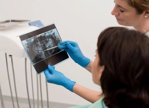
In modern dentistry, accurate diagnosis has become the cornerstone for developing successful treatment plans. This is where contrast imaging plays a vital role as an advanced tool that helps dentists visualize hidden details inside the mouth and jaws. This technique uses special contrast agents to make radiographic images clearer. Dalili Medical helps in detecting complex issues such as root fractures, advanced periodontal disease, bone infections, and even in evaluating the success of dental implants.
Contrast imaging for dentistry is a type of radiographic examination in which a contrast agent (dye) is used to enhance the visibility of internal oral structures, such as root canals, bone cavities, and fine blood vessels.
This technique is usually applied in cases where standard images are insufficient to reveal the problem or when planning precise surgical procedures.
1. Iodine-based contrast
The most commonly used in dental imaging.
Applied with cone-beam computed tomography (CBCT) or conventional CT scans.
Provides high clarity for oral and bone structures.
2. Barium-based contrast
Used in limited cases, such as swallowing assessments or examining soft tissues in the mouth.
Often administered as a mouth rinse solution.
3. Other contrast materials
Such as gadolinium-based agents, used in MRI scans of the jaw or salivary glands.
Diagnosing fine fractures in roots or bones.
Assessing advanced periodontal disease to determine its effect on the bone.
Examining root canals before or after endodontic treatment.
Detecting oral tumors or cysts.
Planning dental implant procedures and determining precise implant locations.
Imaging salivary glands to detect blockages or infections.
1. Cone-Beam Computed Tomography (CBCT) with contrast
Produces a high-resolution, 3D image.
Ideal for surgical planning and dental implant placement.
2. Panoramic X-ray with contrast
Rarely used to highlight specific areas.
Less accurate than CBCT but faster and more affordable.
3. Magnetic Resonance Imaging (MRI) of the jaw
Uses gadolinium-based contrast to visualize soft tissues and salivary glands.
1. Initial assessment
Review the patient’s medical history and ensure there are no allergies to contrast agents or kidney problems.
2. Preparation
Remove any jewelry or removable metal dental work.
3. Administering the contrast
Can be done via intravenous injection or by rinsing with a contrast solution, depending on the exam type.
4. Imaging
Images are taken using a CBCT machine or other suitable radiographic devices.
5. Post-exam
Drink plenty of water to help eliminate the contrast material from the body.
Enhances the visibility of complex details in the mouth.
Facilitates early diagnosis of dental problems.
Improves the accuracy of surgeries and dental implants.
Monitors treatment outcomes and evaluates success.
Although dental contrast imaging is safe in most cases, certain precautions should be taken:
Possible allergic reactions to contrast agents.
Caution in patients with kidney issues.
Temporary sensation of warmth or metallic taste in the mouth after injection.
Inform your dentist of any chronic illnesses or previous allergies.
Drink plenty of water after the examination.
Have the test done in a specialized center with qualified medical staff.
With rapid advancements in medical imaging technology, contrast agents have become safer and less likely to cause complications. New targeted contrast materials are now being developed to focus on specific areas, improving diagnostic accuracy and reducing the need for invasive surgery.
Dental contrast imaging represents an important step toward accurate diagnosis and successful treatment, especially in complex cases that are difficult to detect with conventional imaging. With ongoing medical progress, this technique is expected to become an essential part of modern dental practice, benefiting both dentists and patients alike.


