

In the world of dentistry, X-rays are an essential tool for detecting dental and jaw problems that cannot be seen with the naked eye. While Intraoral Radiographs are the most common type, Extraoral Radiographs play a crucial role in diagnosing complex cases, especially those involving the bones, jaws, and the temporomandibular joint (TMJ).In this article from Dalili Medical, we will take you on a simple and informative journey to understand what Extraoral Radiographs are, their types, uses, benefits, risks, and how to prepare for them.
What Are Extraoral Radiographs?
Extraoral radiographs are a type of dental X-ray in which the film or digital sensor is placed outside the mouth, capturing a larger area that includes the teeth, upper and lower jaws, the temporomandibular joint (TMJ), and surrounding tissues.
This type of X-ray is used when the problem is too large or complex to be clearly seen with intraoral radiographs.
Importance of Extraoral Radiographs
Extraoral radiographs play a key role in:
Diagnosing jaw problems such as fractures or deformities.
Detecting impacted teeth.
Monitoring the growth of teeth and jaws in children.
Locating tumors or cysts within the bone.
Assessing the temporomandibular joint (TMJ) in cases of pain or clicking sounds.
Planning for dental implants or surgical procedures.
Types of Extraoral Radiographs
There are several main types of extraoral radiographs, each serving specific purposes:
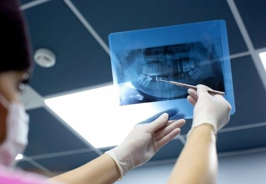
Panoramic Radiograph
Captures the entire dental arch in a single image.
Ideal for detecting impacted teeth, fractures, and gum diseases.
Lateral Cephalometric Radiograph
Used in orthodontics to assess the relationship between the upper and lower jaws and the skull.
Helps in orthodontic treatment planning.
Posteroanterior Cephalometric Radiograph
Shows the face from the front.
Useful for diagnosing skeletal deformities.
TMJ Radiographs
Focuses on the joint connecting the jaw to the skull.
Detects inflammation, dislocation, or degeneration.
Cone Beam Computed Tomography (CBCT)
Provides high-resolution 3D images.
Perfect for surgical planning and dental implant placement.
Steps for Performing Extraoral Radiographs
Preparation
Remove any jewelry or metallic objects from the head and neck area.
Wear a lead apron to protect the body from radiation.
Patient Positioning
The technician positions the patient according to the specific type of X-ray required.
Imaging
The machine operates for just a few seconds.
Image Result
For digital radiographs, the image appears instantly on the screen.
For conventional radiographs, the film is developed before the image can be viewed.
Advantages of Extraoral Radiographs
Cover a wide area of the mouth and jaws.
Detect issues that may be hard to identify with intraoral radiographs.
Ideal for comprehensive treatment planning.
Painless and quick to perform.
Risks of Extraoral Radiographs
Although the radiation dose is relatively low, certain precautions are important:
Wear a lead apron and a thyroid shield.
Avoid exposure during pregnancy unless absolutely necessary.
Use the lowest possible radiation dose.
Differences Between Extraoral and Intraoral Radiographs
| Criterion | Extraoral Radiographs | Intraoral Radiographs |
|---|---|---|
| Sensor Location | Outside the mouth | Inside the mouth |
| Field of View | Includes teeth, jaws, and surrounding tissues | Focuses on a limited number of teeth |
| Usage | Comprehensive diagnosis, jaw and TMJ cases | Detecting cavities and root infections |
| Radiation Dose | Relatively low | Very low |
Recent Advances in Extraoral Radiography
Digital Radiography: Instant, high-quality images with zoom and analysis capabilities.
Cone Beam CT Technology: High-precision 3D imaging.
Low-Dose Machines: Better patient protection.
AI Diagnostic Software: Uses artificial intelligence for early disease detection.
Tips Before Undergoing Extraoral Radiographs
Inform your dentist if you are pregnant.
Remove any metallic objects from your head or neck.
Follow the radiology technician’s instructions carefully.
Try to remain still during imaging for a clear picture.
Conclusion
Extraoral radiographs are an essential tool for diagnosing and treating dental and jaw problems, especially those that are difficult to detect with the naked eye or intraoral radiographs.
With technological advancements, these X-rays have become more accurate and safer, making them an excellent choice for diagnosing complex cases and planning treatments.
If your dentist recommends them, there’s no need to worry—they are quick, painless, and provide valuable diagnostic information.


