

In the world of dentistry, X-rays are an essential tool for detecting problems that cannot be seen with the naked eye. Among the different types of dental X-rays, occlusal radiography holds a special place, as it provides a wide view of the entire upper or lower dental arch. This allows dentists to diagnose complex issues and accurately monitor treatment plans.In this comprehensive guide from Dalili Medical, we will explore the meaning of occlusal radiography, its types, importance, step-by-step procedure, benefits, possible risks, and the latest technologies in this field.
What is Occlusal Radiography?
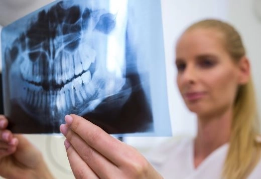
Occlusal radiographs are a type of intraoral X-ray used to capture a complete image of the upper or lower dental arch.
A film or digital sensor is placed inside the mouth on the occlusal surface (where the upper and lower teeth meet), and the X-ray beam is directed from outside at a specific angle to obtain the image.
Occlusal radiography plays a vital role in diagnosing certain conditions that may not be easily detected using other types of X-rays. Its main uses include:
Locating unerupted or impacted teeth.
Diagnosing fractures in the jaws or teeth.
Detecting cysts or tumors in the jaw.
Identifying the location of foreign bodies in the mouth.
Assessing tooth development in children.
Monitoring surgical and orthodontic cases.
1. Maxillary Occlusal
Description: Shows the entire upper arch, including the teeth, palate, and surrounding bone.
Uses:
Locating impacted upper teeth.
Diagnosing cracks or fractures in the upper jaw.
Detecting tumors or cysts.
2. Mandibular Occlusal
Description: Displays the lower arch of teeth and surrounding bone.
Uses:
Locating unerupted lower teeth.
Examining fractures in the lower jaw.
Detecting foreign objects or salivary stones.
3. Lateral Occlusal
Description: Shows one side of the dental arch.
Uses:
Assessing the development of side teeth.
Detecting localized deformities or infections.
1. Preparation
The patient sits upright in the dental chair.
A lead apron is worn to protect the body from radiation.
Any metal objects or jewelry in the head and neck area are removed.
2. Placement of Film or Sensor
The film or digital sensor is placed on the occlusal surface (where the teeth meet).
The patient is instructed to gently close their mouth to hold the film in position.
3. Positioning the X-ray Machine
The device is adjusted to the appropriate angle depending on the required type of occlusal radiograph (upper or lower).
The patient is instructed to remain still and avoid movement.
4. Capturing the Image
The X-ray machine is activated for a few seconds to capture the image.
For digital X-rays, the result appears instantly on the monitor, while traditional films require chemical processing.
Wide Coverage: Captures the entire dental arch in a single image.
Accurate Diagnosis: Ideal for detecting impacted teeth or fractures.
Child-Friendly: Useful for monitoring the development of both primary and permanent teeth.
Relative Safety: Low radiation dose when safety protocols are followed.
Although the radiation dose is low, certain precautions should be taken:
Wearing a lead apron for protection.
Using a thyroid collar when necessary.
Avoiding unnecessary exposure, especially for pregnant patients.
Adjusting the dose according to the patient’s age.
| Criterion | Occlusal X-ray | Bitewing X-ray | Periapical X-ray |
|---|---|---|---|
| Field of View | Entire dental arch | Several adjacent teeth | One or more teeth to the root |
| Common Uses | Impacted teeth, fractures, tumors | Interproximal caries detection | Root infections |
| Radiation Dose | Low | Low | Low |
| Duration | Few seconds | Few seconds | Few seconds |
Digital X-rays: Instant, high-quality images with zoom capability.
Diagnostic Software: Image analysis for early detection of problems.
Low-Radiation Devices: Significantly reduce patient exposure.
3D Technology: Combining occlusal radiographs with cone beam CT for enhanced imaging.
Inform your dentist if you are pregnant.
Remove any metallic objects from your head or mouth.
Stay completely still during the procedure.
Follow the dentist’s or radiographer’s instructions.
Occlusal radiographs are an essential diagnostic tool in dentistry, offering a comprehensive view of the dental arch and helping to identify problems that may not be detected by other imaging methods. With advances in technology, these X-rays are now more accurate, safer, and faster, making them a valuable choice for both diagnosis and treatment follow-up.


