

Have you ever felt a deep pain in one of your teeth without knowing the cause? The problem might be hidden in the tooth’s root or the surrounding bone—areas that cannot be seen with the naked eye or even during a routine dental checkup. This is where the Periapical X-ray comes in. It is an essential tool that gives the dentist a complete view of the tooth from crown to root, along with the surrounding bone, to detect infections, cysts, or any hidden issues. In this article from Dalili Medical, we’ll take you on a simple, informative journey to understand everything you need to know about Periapical X-rays: what they are, when you might need them, how they are performed, whether they are safe, and tips to keep your teeth healthy.
What is a Periapical X-ray?
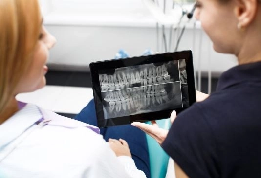
A Periapical X-ray is a type of intraoral dental X-ray used to capture a complete image of a single tooth or a small group of teeth—from the crown to the root tip—along with part of the surrounding bone.
It is called periapical because the “apex” refers to the end of the tooth’s root, and this X-ray focuses on imaging that area in detail to detect any issues.
When Does a Dentist Recommend a Periapical X-ray?
A dentist may request this type of X-ray in the following cases:
Diagnosing infections or abscesses at the tooth root.
Detecting cysts or tumors in the jaw area.
Assessing tooth injuries after accidents or trauma.
Monitoring the success of root canal treatment.
Examining teeth before dental implant placement.
Evaluating bone condition before crowns, bridges, or dentures.
Identifying impacted or hidden teeth problems.
Steps of a Periapical X-ray Procedure
Patient Preparation
Wearing a lead apron to protect the body from radiation.
Explaining the procedure to the patient to reduce anxiety.
Placing the Film or Digital Sensor
The film or sensor is placed inside the mouth near the target tooth.
The patient is asked to hold it gently in place.
Taking the X-ray
The X-ray machine is carefully positioned towards the target area.
The image is captured within seconds.
Reviewing the Results
The image is displayed on a computer screen (for digital X-rays) or on traditional X-ray film.
The dentist interprets the results and decides on the appropriate treatment plan.
Provides a complete image of the tooth from crown to root.
Accurately detects root and surrounding bone problems.
An essential tool for root canal treatment.
Helps in planning for dental implants or surgeries.
Relatively safe with a low radiation dose.
Yes, they are generally safe, especially when using lead aprons and modern digital X-ray techniques that reduce radiation exposure.
The amount of radiation from a single image is roughly equivalent to what a person receives naturally from the environment in one or two days.
⚠️ Note: X-rays should be limited to when they are medically necessary, particularly for pregnant women in their first trimester.
When symptoms like pain or swelling are present.
Before starting root canal treatment or dental implant placement.
When a cyst or abscess is suspected.
To monitor the condition of treated teeth.
| Type of X-ray | What It Shows | Main Use |
|---|---|---|
| Periapical X-ray | Entire tooth from crown to root + surrounding bone | Diagnosing root and bone problems |
| Bitewing X-ray | Crowns of upper and lower teeth in the same area | Detecting cavities and gum disease |
| Panoramic X-ray | Full image of jaws and teeth | Comprehensive evaluation before surgery or implants |
| Occlusal X-ray | All teeth in one jaw | Checking growth patterns or abnormalities |
A dentist may request periapical X-rays for children in cases such as:
Injuries to permanent or baby teeth after a fall or impact.
Suspected abscesses or infections.
Monitoring tooth development or detecting root problems.
No special preparation is needed.
Brushing teeth before the appointment is recommended.
Inform the dentist if you are pregnant.
Remove any metallic jewelry from the face area.
Digital X-rays: Provide instant, high-resolution images with the ability to zoom and adjust.
3D Imaging (CBCT): Offers highly detailed views of roots and surrounding bone, especially useful for complex surgical cases.
Costs vary depending on the country and equipment used:
Egypt: 50–150 EGP per image.
Gulf countries: 20–50 SAR.
Europe & USA: 10–30 USD.
1. Are Periapical X-rays painful?
No, the procedure is painless, though you may feel slight discomfort from the device inside your mouth.
2. How long does the procedure take?
Usually less than 5 minutes.
3. Can children have this type of X-ray?
Yes, using special sizes designed for a child’s mouth.
4. Are there alternatives?
Panoramic X-rays or 3D imaging can provide similar information, but periapical X-rays offer the most precise view of the tooth roots.
Periapical X-rays are an indispensable diagnostic and treatment tool for detecting and managing deep dental issues, especially those involving tooth roots and surrounding bone. Thanks to their accuracy and ease of use, they enable dentists to identify problems early and provide effective treatment, preserving oral health in the long term.
Remember: Regular check-ups and visiting your dentist at the first sign of pain can save your teeth from loss and prevent the need for complex treatments.


