

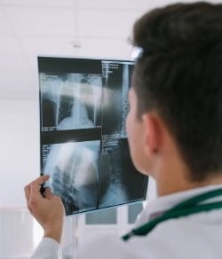
PET/MRI is an advanced imaging technique that combines two types of scans:
PET (Positron Emission Tomography): Used to track the biological activity of cells inside the body.
MRI (Magnetic Resonance Imaging): Provides detailed images of tissues and organs using magnetic fields and radio waves.
By merging these two technologies, PET/MRI produces highly detailed images that clearly show both the anatomical structure and cellular activity simultaneously. This makes PET/MRI one of the most powerful modern diagnostic tools.
PET/MRI is applied in several advanced medical fields, including:
Accurate Tumor Diagnosis
Differentiates between benign and malignant tumors.
Monitors cancer spread and treatment response.
Brain and Nervous System Disorders
Diagnoses brain tumors, epilepsy, and neurodegenerative diseases like Alzheimer’s and Parkinson’s.
Assists in precise surgical planning.
Heart Diseases
Evaluates blood flow to the heart muscle and detects damaged tissue.
Personalized Medicine Research
Helps determine effective treatments based on individual responses.
PET/MRI offers clearer, more accurate images of soft tissues (such as the brain, liver, and reproductive organs).
PET/CT is faster and slightly less expensive but exposes patients to higher radiation due to the CT scan.
PET/MRI is often preferred when imaging sensitive areas or needing detailed soft tissue analysis.
Preparation
Patients are usually asked to fast for 4 to 6 hours before the scan.
Inform medical staff if you are pregnant, breastfeeding, have allergies, or have implanted medical devices like pacemakers.
Injection of Radioactive Tracer
A safe radioactive substance called FDG (similar to glucose) is injected.
You wait 30 to 60 minutes for it to circulate.
Scanning Procedure
You lie down on a bed that slowly moves into the PET/MRI machine.
The scan lasts from 45 minutes to 90 minutes.
Immobility During Scan
It is important to remain still to ensure clear images.
✅ Completely painless.
✅ Relatively safe, with only a minor sting when injecting the tracer.
Precautions:
Not recommended for pregnant or breastfeeding women unless absolutely necessary.
Patients with claustrophobia may require mild sedation.
Inform your doctor if you have metal implants.
Extremely high accuracy in detecting tumors and cellular activity.
Ideal for brain and spinal cord imaging.
Reduced radiation dose compared to PET/CT.
Combines anatomical and functional images simultaneously.
Supports personalized medicine and precise treatment planning.
⏳ Longer scan times compared to other imaging methods.
Higher cost than alternative imaging techniques.
Limited availability in some hospitals and medical centers.
Q: Does PET/MRI expose me to harmful radiation?
A: It does involve radiation, but at a very low dose compared to PET/CT and is considered safe for medical use.
Q: How long does the scan take?
A: Approximately 45 to 90 minutes, depending on the area examined.
Q: Can I drive after the scan?
A: Yes, unless you were given sedation, in which case you should have someone accompany you.
This depends on your medical condition. PET/MRI is ideal if your doctor needs the most accurate anatomical and functional imaging—especially for tumors or neurological disorders.
The PET/MRI scan is one of the most advanced imaging techniques globally, combining the detailed anatomical imaging of MRI with PET’s ability to show cellular activity. It helps diagnose complex diseases with high accuracy while minimizing radiation exposure compared to other technologies. Although it is relatively costly, its diagnostic value can significantly influence your treatment plan.


