

Have you been asked to undergo a SPECT scan?
Have you heard about this technique and wondered how it helps in diagnosing diseases? In this article from Dalily Medical, we provide you with a simple and comprehensive guide to understanding SPECT imaging (Single Photon Emission Computed Tomography) — what it is, how it's done, its key medical uses, and how it differs from other tests like PET scans or traditional CT scans.
SPECT stands for Single Photon Emission Computed Tomography.
It is a type of nuclear medicine imaging that provides 3D images of blood flow and organ function inside the body. This is achieved by tracking a small amount of radioactive material injected into a vein.
Blood flow to the heart or brain
Organ activity and function
Detection of tumors, infections, or seizure activity
Although both use radioactive tracers, there are key differences:
| Factor | SPECT | PET Scan |
|---|---|---|
| Radioactive substance | Photon emitters | Positron emitters |
| Cost | Lower | Higher |
| Image precision | Good | Higher precision |
| Common uses | Heart, brain, bones | Cancer, brain, heart |
Summary: PET scans offer higher image clarity but are more expensive. SPECT is more widely available and cost-effective.
Doctors use SPECT to help diagnose and monitor various conditions:
Detect reduced blood flow (ischemia)
Evaluate the effectiveness of bypass surgery or stents
Identify damaged or dead heart tissue post-heart attack
Diagnose and locate seizure activity (epilepsy)
Evaluate blood flow after a stroke
Assist in diagnosing Alzheimer’s or dementia
Check cancer spread in bones or organs
Monitor response to chemotherapy or radiation
You may need to fast or avoid certain medications.
Wear comfortable clothes and remove any metal (earrings, necklaces, etc.).
A small dose of radioactive tracer (commonly Technetium-99m) is injected into a vein.
You’ll wait a while (minutes to hours) for the tracer to distribute in the body.
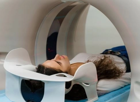
You’ll lie on a table that slides into a scanner similar to a CT machine.
The camera rotates around your body to collect multiple images that are later reconstructed into 3D visuals.
Varies by organ being scanned, typically 30 to 60 minutes.
✅ Yes, it is generally safe.
The amount of radiation used is low and within safe limits for most people.
Exceptions:
Not recommended for pregnant or breastfeeding women unless absolutely necessary.
Mild discomfort at injection site
Temporary nausea or headache
Slight fatigue
Inform your doctor if you are pregnant or breastfeeding.
Let them know if you have any known allergies to radioactive materials.
Follow instructions regarding fasting or medication.
Wear loose clothing without metal and avoid jewelry or watches.
Normal results: Normal blood flow and organ function.
Abnormal results: May indicate blocked arteries, brain abnormalities, tumors, or inflammation.
A radiologist interprets the images and sends the results to your doctor, who will explain them and plan next steps.
Is the scan painful?
No. It is painless, except for a small prick when injecting the tracer.
How long does the tracer stay in my body?
The radioactive material typically leaves your system within 24 to 48 hours. Drinking water helps speed up elimination.
Can I drive after the scan?
Yes. Most patients can return to normal activities immediately.
Absolutely. A SPECT scan is a safe, non-invasive, and highly informative tool that gives doctors valuable insights into organ function — information that traditional imaging cannot provide.
If your doctor recommends a SPECT scan, don’t worry. Think of it as a smart step toward accurate diagnosis and effective treatment planning.


