

Has your doctor recommended a thyroid imaging test using ultrasound or radioactive isotopes? Don’t worry—these are safe, accurate diagnostic tools that help physicians evaluate the condition of your thyroid gland and determine the cause of symptoms such as:
Neck swelling (goiter)
Nodules or lumps in the thyroid
Hormonal imbalances (such as hyperthyroidism or hypothyroidism)
Monitoring thyroid cancer or post-surgical follow-up
In this article from Dalili Medical, we’ll walk you through a complete and simple guide to thyroid imaging: the types of scans available, the difference between ultrasound and nuclear imaging, when each test is recommended, how to prepare, and any potential risks.
Types of Thyroid Imaging: What You Need to Know
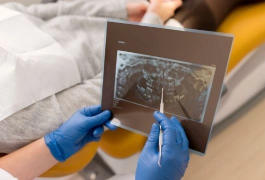
Thyroid ultrasound is the most commonly used imaging test to evaluate the thyroid gland. It uses high-frequency sound waves to produce real-time images of the thyroid.
✅ Benefits of Ultrasound:
Completely painless
No radiation exposure
Quick (10–15 minutes)
Shows thyroid size, nodules, and calcifications
❓When Is It Used?
Detecting thyroid nodules and determining if they’re solid or fluid-filled
Monitoring changes in nodules over time
Guiding a fine needle aspiration biopsy (FNA) for suspicious nodules
This test uses radioactive isotopes (such as Iodine-123 or Technetium-99m) taken orally or injected. A special gamma camera captures images of the thyroid after the tracer is absorbed.
✅ What It Shows:
Functional activity of different thyroid regions (active, underactive, or normal)
Differentiates between:
“Hot” nodules: absorb the isotope and produce hormone (often benign)
“Cold” nodules: do not absorb the isotope (may require further evaluation)
Helps assess hyperthyroidism or an enlarged thyroid (goiter)
❗ Important: Nuclear scans are not typically used in pregnant, breastfeeding, or young pediatric patients unless absolutely necessary and under special guidance.
| Feature | Ultrasound | Nuclear Scan |
|---|---|---|
| Type of Exam | Anatomical (structure) | Functional (activity) |
| Radiation | None | Low-dose radioactive material |
| Duration | 10–15 mins | 30–60 mins |
| Used For | Shape, size, and nodules | Gland activity and hot/cold nodules |
| Pregnancy Safe? | Yes | No |
No special preparation required
Eat and take medications normally
Remove necklaces or metal around the neck
Inform your doctor if you:
Are pregnant or breastfeeding
Are taking thyroid medications (like levothyroxine)
Recently had iodine-based tests or consumed iodine-rich foods
You may need to:
Stop certain medications for a few days
Fast for several hours
Avoid iodine-rich food before the scan
Nuclear scan shows increased uptake in all or part of the thyroid (e.g., in Graves’ disease or toxic nodules).
Scan shows reduced isotope absorption, indicating poor gland activity.
Ultrasound reveals the size, shape, and nature of nodules.
Nuclear scan indicates if nodules are functioning ("hot") or inactive ("cold").
Nuclear imaging helps detect remaining thyroid tissue or cancer cells after thyroid surgery or cancer therapy.
Completely safe, non-invasive, and no side effects.
Low radiation dose, generally safe for most adults.
You may be advised to avoid close contact with children and pregnant women for 24 hours post-scan.
Rare allergic reactions may occur to the tracer.
Can both tests be done together?
Yes. In some cases, both ultrasound and nuclear scans are used to give a complete view of the thyroid’s structure and function.
Does ultrasound replace nuclear scanning?
Not always. Ultrasound is ideal for examining the structure, but nuclear scanning gives insights into thyroid function.
Is the nuclear scan painful?
No. You may feel a small pinch during the injection or a slight taste in your mouth if taken orally.
Whether you're dealing with thyroid nodules, symptoms of hyperthyroidism, or post-surgical follow-up, thyroid ultrasound and nuclear scans are key diagnostic tools. Each test plays a unique role—ultrasound shows structure, while nuclear imaging reveals function.


