

Do you suffer from joint, knee, or shoulder pain? Has your doctor recommended an imaging scan for these areas? Don’t worry—medical imaging has become one of the most essential tools for accurately diagnosing bone and joint problems without the need for surgery.In this article from Dalilimedical, we offer you a comprehensive guide to joint, knee, and shoulder imaging. You'll learn about the different types of scans, when they're used, what they reveal, and how to prepare for them.
Imaging tests are essential for clearly visualizing bones and joints. They help in:
Identifying the cause of pain or swelling
Detecting joint inflammation
Evaluating fractures or dislocations
Diagnosing arthritis or ligament injuries
Monitoring chronic conditions like rheumatoid arthritis
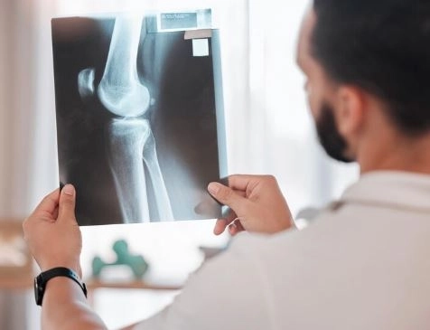
The most commonly used scan for bone assessment
Show fractures, dislocations, and joint space narrowing due to osteoarthritis
Frequently used for the knee, shoulder, and ankle
Offers detailed images of soft tissues (ligaments, cartilage, tendons)
Ideal for diagnosing:
ACL tears and meniscus injuries in the knee
Rotator cuff injuries in the shoulder
Joint inflammation and fluid accumulation
Used when X-rays are not detailed enough
Provides 3D cross-sectional images
Excellent for complex fractures or post-traumatic joint injuries
Evaluates soft tissue swelling, joint fluid, or tendon tears
Quick, safe, and radiation-free
Commonly used for shoulder, knee, or hand joints
Knee imaging helps diagnose:
Knee osteoarthritis
Joint infections or inflammation
ACL or meniscus tears
Swelling or fluid in the joint
Sports injuries or trauma-related conditions
The shoulder is a complex joint that requires detailed imaging for accurate diagnosis. Scans may reveal:
Tendonitis or tendon tears
Recurrent shoulder dislocations
Rotator cuff injuries
Joint calcifications or degeneration
Bursitis (inflammation of the fluid-filled sacs around the joint)
Imaging isn’t limited to the knee and shoulder. It’s also used for:
Hand and finger joints: Especially to diagnose rheumatoid arthritis
Ankle: To assess sprains or fractures
Hip and pelvic joints: Common in elderly patients or after a fall
Most joint scans require little to no preparation:
Wear comfortable clothing and avoid metal accessories (jewelry, pins)
Inform your doctor if you are pregnant before undergoing any radiation-based scan
For MRI, notify your doctor if you have a metal implant or pacemaker
| Imaging Type | Approximate Duration |
|---|---|
| X-ray | 5–10 minutes |
| MRI | 30–60 minutes |
| CT Scan | 10–20 minutes |
| Ultrasound | 15–30 minutes |
After your scan, your doctor will provide a detailed report, which includes:
A description of findings (e.g., arthritis, tear, swelling)
Recommendations for further tests or follow-up
Suggested treatment options (medication, physiotherapy, or surgery)
Is joint imaging painful?
No, all imaging procedures are completely painless.
Can multiple imaging types be done on the same joint?
Yes. Sometimes a doctor may request more than one type for a more accurate diagnosis.
How often can joint imaging be repeated?
It depends on the type:
X-rays and CT scans involve radiation, so they should be limited
MRI and ultrasound are safe and can be repeated when necessary
Joint, knee, and shoulder imaging plays a vital role in diagnosing musculoskeletal issues accurately. The type of imaging used depends on your symptoms and your doctor’s evaluation.
Always feel free to ask your doctor any questions before the scan to ensure you're fully comfortable and understand your condition clearly.
Remember: early diagnosis leads to faster treatment and better outcomes.


