

Has your doctor recommended an echocardiogram? Are you wondering what this test is and why it's commonly requested?Don’t worry—this article from Dalili Medical offers you a simplified yet comprehensive guide to echocardiography, also known as heart ultrasound. We’ll walk you through everything you need to know about this important test, its types, why it's done, and what to expect during the procedure.
Echocardiography, commonly known as heart ultrasound, is a non-invasive diagnostic test that uses ultrasound waves to create real-time images of the heart. It helps assess the heart's size, shape, pumping efficiency, valve function, and detect abnormalities in cardiac structure or performance.
This test is very similar to the ultrasound used during pregnancy but focuses on the heart, providing live images of the heartbeat and blood flow.
Echocardiograms are essential tools in diagnosing many heart conditions. A doctor may request this test in the following situations:
Chest pain or palpitations
Unexplained shortness of breath
Suspected heart failure
Evaluation of heart valve diseases
Monitoring of chronic heart conditions
After a heart attack
Pre-operative assessment before major surgeries
Diagnosing congenital heart defects in children
There are several types of echocardiograms, and the choice depends on the condition being evaluated:
Transthoracic Echocardiogram (TTE)
The most common type
A transducer is placed on the chest to produce heart images
Transesophageal Echocardiogram (TEE)
A probe is inserted through the mouth into the esophagus
Provides clearer images of heart valves and detects clots in the left atrium
Stress Echocardiography
Done during or after physical exercise (or using medication)
Assesses heart function under stress
3D/4D Echocardiography
Produces highly detailed 3D images of heart chambers and valves
Often used for surgical planning or post-surgical assessment
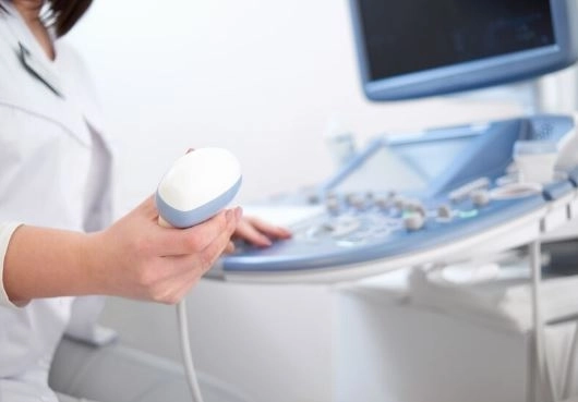
Echocardiography is simple and usually takes 20–40 minutes. Here's how it works:
Preparation:
No fasting is required for most types, except for TEE (requires 6-hour fasting).
Positioning:
You’ll lie on an exam table, usually on your left side.
Imaging:
A technician applies gel to the chest and uses a handheld device (transducer) to capture heart images.
Analysis:
Images are displayed in real-time and later interpreted by a cardiologist.
An echocardiogram helps detect or assess the following:
Strength of heart muscle contraction
Enlargement or weakness of the heart
Valve disorders (stenosis, regurgitation)
Pericardial effusion (fluid around the heart)
Congenital heart defects
Blood clots inside heart chambers
Cardiac tumors
Pulmonary hypertension (high pressure in the lung artery)
Non-invasive and painless
No harmful radiation
Safe for all age groups, including pregnant women
High accuracy in assessing heart function
Can be repeated for ongoing monitoring
Yes, echocardiography is completely safe and well-tolerated by patients of all ages.
Unlike X-rays or CT scans, it does not involve radiation. Only TEE may cause slight temporary discomfort due to the probe placement.
| Feature | Echocardiogram | ECG (Electrocardiogram) |
|---|---|---|
| Type of Test | Ultrasound Imaging | Electrical Activity Recording |
| Purpose | Heart structure, function, valves | Heart rhythm and electrical signals |
| Duration | 20–40 minutes | 5–10 minutes |
| Usage | Structural and functional diagnosis | Rhythm monitoring and arrhythmias |
Does echocardiography detect clots?
✅ Yes. Especially TEE can detect small clots in the left atrium or ventricle.
Can echocardiography show blocked arteries?
❌ Not directly. However, it may indicate damage to the heart muscle caused by reduced blood flow. Stress echo is used in such cases, and follow-up with CT angiography or cardiac catheterization may be required.
Is fasting required for an echocardiogram?
❌ Not usually. Only TEE requires fasting (6 hours before the test).
| Type of Echocardiogram | Estimated Price Range (EGP) |
|---|---|
| Transthoracic Echo | 700 – 1200 EGP |
| Stress Echocardiography | 1200 – 2000 EGP |
| Transesophageal Echo | 1500 – 2500 EGP |
| 3D/4D Echocardiography | 2000 – 3000 EGP |
Note: Prices vary by clinic, location, and urgency of results.
Echocardiography is one of the most essential, safe, and accurate tools for diagnosing heart conditions, including valve diseases, heart muscle performance, and congenital defects. It’s non-invasive, widely available, and plays a crucial role in early detection and treatment planning.
If your doctor recommends an echocardiogram, there's no need to worry—it’s a simple and valuable step toward protecting your heart health.


