

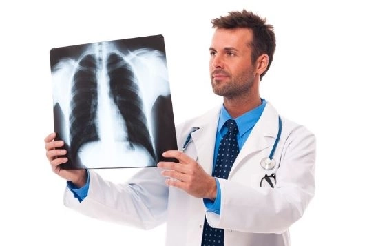
Bronchoscopy is an important medical procedure used by doctors to diagnose and treat respiratory problems, especially those affecting the lungs and the trachea. This procedure helps visualize the airways from the inside using a thin, flexible tube equipped with a small camera. It can reveal many causes behind persistent cough, shortness of breath, or the presence of blood in the sputum.In this Dailly Medical article, we’ll explore together the purpose of bronchoscopy, the steps involved in the procedure, the main potential risks, and the expected outcomes—so you can have a clear understanding before undergoing it yourself or if someone close to you needs it.
What Is Bronchoscopy?
Bronchoscopy is a simple medical procedure performed by a doctor to view the airways and lungs from the inside using a thin tube equipped with a small camera and light, known as a bronchoscope. This procedure helps diagnose issues such as persistent cough, blood in the sputum, or difficulty breathing. It can also be used to treat certain conditions.
There are two main types of bronchoscopes:
Flexible bronchoscope: This is the most commonly used type. It’s thin and can move easily inside the airways. Doctors often use it to take tissue samples (biopsies), suction secretions, or check if the airway is clear.
Rigid bronchoscope: This one is less flexible and more robust. It's used for more complex procedures, such as removing large foreign objects, controlling bleeding, or placing stents to treat tumors.
In most cases, patients receive anesthesia—either general anesthesia to make them fully asleep, or local anesthesia to numb only the area being examined. The type of anesthesia depends on the patient’s condition and the type of bronchoscope being used.
Bronchoscopy usually takes between 30 to 90 minutes, depending on the reason for the procedure. If it’s a simple diagnostic exam, it may be done quickly. But if the doctor needs to remove something or take samples, it may take longer.
Generally, bronchoscopy is very safe. However, like any medical procedure, there are a few rare risks. One of these is lung collapse (also known as atelectasis), which can occur, especially if general anesthesia is used. But this is very rare, and doctors always take the necessary precautions to reduce any potential risks.
The main goal is to make you feel as comfortable as possible. If local anesthesia is used, you might feel some discomfort or a strange sensation as the tube passes through your mouth or nose—but it’s not usually painful. If you’re under general anesthesia, you won’t feel anything at all during the procedure.
For children, doctors usually use a flexible bronchoscope, as it’s safer and more suitable for smaller airways. The procedure is typically done under general anesthesia so the child stays still and comfortable throughout. In most cases, it lasts about 20 to 45 minutes, depending on the child’s condition and the purpose of the bronchoscopy.
Bronchoscopy is a highly effective tool for examining and diagnosing various respiratory issues, such as:
Lung cancer: One of the most common cancers worldwide, detected in millions of patients annually.
Infections: Including pneumonia and tuberculosis, which affect a large number of people each year.
Chronic Obstructive Pulmonary Disease (COPD): A long-term condition that severely affects breathing and impacts millions globally.
Interstitial lung diseases: These cause damage to lung tissues.
Unexplained chronic cough: When someone has a persistent cough with no clear reason, bronchoscopy can help uncover the cause.
Recovery usually takes at least 45 minutes, and sometimes a bit longer depending on the individual. During this time, healthcare staff will monitor your vital signs and ensure everything is stable and there are no unusual side effects.
After bronchoscopy, you should wait until the numbness in your throat wears off—this may take an hour or more. It’s very important not to eat or drink too soon to avoid accidentally inhaling food or liquids into the lungs, which could lead to choking or aspiration. Once the numbness is gone, start with small sips of water and light, easy-to-digest foods.
For simple procedures, the doctor may use local anesthesia to numb part of the throat so your child stays comfortable. However, in more complex cases, especially those done in hospitals, children usually receive general anesthesia to stay asleep throughout the procedure. This is done under the care of an anesthesiologist and a trained medical team to ensure the child’s safety.
A bronchoscopy is a medical procedure that allows doctors to view the inside of the airways and lungs—especially the bronchi and smaller bronchioles. Here's a step-by-step look at how it works:
Before the procedure, you or your child may be given a sedative to help relax. A local anesthetic is also applied to numb the back of the throat. In some cases, general anesthesia may be needed depending on the situation.
The bronchoscope is a thin, flexible tube with a tiny camera and light at the end. The doctor inserts it through the mouth or nose—or sometimes through a small opening in the trachea if one exists. You may be asked to swallow gently to help guide the scope.
As the scope moves through the airways, it sends live video to a monitor, allowing the doctor to examine the lungs in detail and identify any abnormalities.
During the procedure, the doctor may collect samples of tissue, mucus, or fluid for lab testing. This helps in diagnosing infections, inflammation, or tumors.
In some cases, a small piece of lung tissue (biopsy) may be taken for a more detailed analysis.
Bronchoscopy can also be therapeutic—not just diagnostic. It may be used to:
Remove a foreign object
Suction out mucus
Place stents to open blocked airways
After it's done, you'll be monitored for a short time to ensure everything is okay—especially if sedation or anesthesia was used. This helps ensure your safety and supports quick recovery.
Doctors use bronchoscopy for a variety of reasons to diagnose or treat lung and airway conditions. Here are the most common:
If you’ve had a persistent cough for weeks with no clear cause, bronchoscopy helps identify whether it’s due to inflammation, infection, a foreign object, or a tumor.
If blood appears in your mucus when you cough, bronchoscopy can help locate the bleeding source—whether from infection or tumors.
If a CT scan or chest X-ray shows a mass or spot in the lungs, a bronchoscopy allows the doctor to take a biopsy from that area for analysis.
In complex infections like tuberculosis or fungal infections, bronchoscopy helps collect samples from inside the lungs to determine the exact type of infection.
Diseases like pulmonary fibrosis or sarcoidosis often require tiny tissue samples from the lungs, which can be taken during a bronchoscopy.
Especially common in children or the elderly, foreign bodies that accidentally enter the airway can be safely removed via bronchoscopy.
Blockages can be caused by tumors, chronic inflammation, or scarring. Bronchoscopy helps locate the problem and plan the treatment.
Before lung or airway surgery, doctors may perform bronchoscopy to assess the condition of the respiratory system.
Bronchoscopy isn’t just for diagnosis. It can:
Remove thick secretions
Dilate narrowed airways using a balloon
Place stents to keep airways open
Bronchoscopy allows doctors to view inside the lungs using a camera-equipped tube. There are two main types, and each serves specific purposes based on the patient’s condition and treatment goals:
This is the most commonly used type, typically performed under mild sedation or local anesthesia.
Advantages:
Minimally painful and more comfortable
Inserted through the nose or mouth
Suitable for both examination and sample collection
Can remove secretions or foreign bodies
Used For:
Diagnosing various lung diseases
Detecting tumors
Monitoring chronic infections
Investigating unexplained bleeding
This is the most commonly used type and is usually done under local anesthesia or mild sedation.
Advantages:
Less painful and more comfortable for the patient
Inserted through the nose or mouth
Suitable for examination, sample collection (biopsies), and mucus removal
Used for:
Diagnosing various lung diseases
Detecting tumors
Monitoring chronic infections
Investigating unexplained bleeding
Evaluating chronic cough and shortness of breath
An older, larger instrument that requires general anesthesia, used in more complex or emergency situations.
Advantages:
Allows for more advanced therapeutic procedures
Better control of bleeding
Removal of large foreign objects
Insertion of stents to keep airways open
Used for:
Airway blockage due to tumors
Removing solid foreign bodies
Controlling severe lung bleeding
A non-invasive digital imaging technique that uses 3D CT scans to create a virtual view of the airways.
Advantages:
Non-surgical and requires no anesthesia
Useful for surgery planning and follow-up
Limitations:
Cannot collect samples or perform treatments
An advanced form of flexible bronchoscopy that includes an ultrasound probe at the tip of the scope.
Advantages:
Provides detailed images of lymph nodes and deep lung areas
Allows sampling of difficult-to-reach areas without surgery
Used for:
Diagnosing and staging lung cancer
Evaluating enlarged lymph nodes
Diagnosing conditions like tuberculosis or sarcoidosis
A modern technique using a GPS-like system to guide the bronchoscope deep into the lungs.
Advantages:
Access to hard-to-reach lung areas
High accuracy in sampling small peripheral tumors
Used for:
Diagnosing small or unclear tumors
Taking precise biopsies from deep lung tissues
Combines bronchoscopy with advanced therapeutic procedures like tumor removal or airway widening.
Used for:
Treating airway tumors
Controlling bleeding
Dilating narrowed airways with stents or balloons
A specially designed bronchoscopy for children, with smaller instruments suited for their anatomy.
Used for:
Diagnosing congenital airway abnormalities
Removing foreign objects like coins or toys
Evaluating chronic cough and recurrent infections
Uses real-time X-ray imaging (fluoroscopy) to guide the bronchoscope accurately inside the lungs.
Advantages:
Pinpoint accuracy for tissue biopsies
Very useful in detecting and reaching small or hidden tumors
Used for:
Diagnosing tumors not easily visible
Precisely directing medical instruments within the lung
Uses special light filters to enhance the visibility of blood vessels and subtle changes in airway linings.
Advantages:
Detects abnormal tissue changes with high precision
Helps identify early-stage cancers
Used for:
Early lung cancer detection
Assessing the effectiveness of chemotherapy or radiation
Uses a special light that makes healthy and abnormal tissues appear in different colors.
Advantages:
Highly sensitive in detecting early cancerous cells
Can reveal changes that are not visible in standard bronchoscopy
Used for:
Monitoring patients after lung cancer treatment
Detecting pre-cancerous changes
A procedure done during bronchoscopy where a saline solution is flushed into a part of the lung and then collected for analysis.
Advantages:
Provides detailed information about infections, inflammation, or abnormal cells
Used for:
Diagnosing various lung infections
Identifying immune or allergic lung diseases
Detecting lung cancer
The most advanced form of bronchoscopy where a doctor controls a robot to navigate very precisely inside the lungs.
Advantages:
Extremely accurate navigation
Safely reaches difficult and deep lung areas
Used for:
Detecting and treating small peripheral tumors
Collecting highly precise biopsy samples with minimal risk
Bronchoscopy Diagnosis and Procedure: A Step-by-Step Easy Guide
Bronchoscopy is an important medical procedure used to examine the lungs and airways from the inside. It helps in both diagnosing and treating various health conditions. The procedure is performed using a thin, flexible tube equipped with a camera and light, which is inserted through the nose or mouth into the airways.
You’ll usually need to fast (avoid food and drink) for a few hours before the procedure.
Your doctor will review your medical history and current medications.
A sedative or local anesthetic may be given to keep you comfortable during the procedure.
The bronchoscope is gently inserted through the nose or mouth into the airway.
The camera on the scope shows clear images of the airways and lungs on a screen, allowing the doctor to detect any abnormalities.
The doctor may take tissue samples (biopsies), mucus, or fluids for lab testing.
If the doctor spots abnormal areas, tissue samples may be collected and examined under a microscope.
Biopsies are essential to diagnose conditions like lung cancer, infections, or other lung diseases.
A saline solution may be used to wash parts of the lung, and a small brush can be used to collect cell samples.
These samples help detect infections, inflammation, or abnormal cells.
Besides diagnosis, bronchoscopy can also be used to:
Remove foreign objects
Suction out secretions
Place stents to open blocked airways
Control bleeding in some cases
After the procedure, you’ll rest under observation until the effects of sedation wear off.
You might feel a mild sore throat, a light cough, or notice a bit of bleeding — this is usually normal.
Samples taken during the procedure are sent to a lab for analysis.
Based on the results, the doctor will give you a clear diagnosis and the best treatment plan.
You may still feel the effects of sedation and not remember the procedure.
You'll stay in the hospital or exam center for a few hours for monitoring and to make sure there are no complications.
It’s common to feel numbness or tingling in your mouth or throat for a few hours.
During this time, avoid eating or drinking hot or solid foods to prevent choking.
Your doctor will start you off with sips of water.
Then, you can move on to light, soft foods like soup or mashed meals.
A mild sore throat or hoarseness may last for a few days.
A light cough is also a normal reaction.
Contact your doctor right away if you notice:
Sudden trouble breathing
Coughing up blood
Severe or lasting chest pain
While bronchoscopy is usually safe, it's not suitable for everyone. Some people may face higher risks, such as:
If you have serious breathing problems or very low oxygen levels, the procedure might be too risky.
People with clotting problems (e.g., hemophilia) have a higher chance of bleeding during or after the procedure.
If you suffer from severe heart disease, your doctor might avoid doing the procedure due to the stress it may cause.
If you've recently had a heart attack or stroke, bronchoscopy is usually postponed until your health stabilizes.
High, uncontrolled blood pressure raises the risk of complications during the procedure.
If you have a severe lung or airway infection, the procedure may be delayed until you're better.
If you’ve had allergic reactions to sedatives or anesthetics in the past, inform your doctor so they can adjust the plan.
Although bronchoscopy is generally safe, complications can occur:
Bleeding: Minor bleeding may occur if a biopsy is taken. Rarely, it can be heavy and require immediate care.
Infection: Lung infections are rare but possible, especially in people with weak immune systems. Some doctors prescribe preventive antibiotics.
Bronchospasm (Airway Spasm): May cause temporary breathing difficulty.
Lung or Airway Injury: Very rarely, the scope can puncture the lung or airway, leading to a collapsed lung (pneumothorax).
Low Oxygen Levels: Oxygen might temporarily drop during the procedure and will be monitored.
Sedation Reactions: Dizziness, nausea, or allergic reactions can happen, but are usually mild.
Hoarseness or Voice Loss: The scope might irritate the vocal cords, causing temporary hoarseness.
Persistent Coughing: A strong cough may occur but usually fades with time.
Sore or Burning Throat: This discomfort can last a few hours to a day and improves with warm fluids.
Low Blood Pressure or Fast Heart Rate: These may be caused by stress or sedative drugs and are carefully monitored.
Temporary Airway Blockage: Swelling or narrowing may occur, requiring hospital observation.
Bronchoscopy is usually performed by a pulmonologist, a doctor specialized in lung and airway conditions.
In some cases, especially when surgery is involved, a thoracic surgeon or an anesthesiologist may also be part of the team to manage anesthesia.


