

Have you ever noticed sudden swelling in your face or unexplained puffiness in your legs? The reason might be the vena cava, one of the most important veins in the body. It plays a vital role in returning blood from the body back to the heart.In this Daleely Medical article, we’ll explore the difference between the superior and inferior vena cava, and highlight the key symptoms that may appear when either one is blocked or affected. We’ll also explain the main diagnostic methods and treatment options—whether through medication or surgery.If you care about your health and want to understand your body better, this article will be your ultimate guide.
What Is the Vena Cava?
The vena cava is the largest vein in the human body. Its main function is to carry deoxygenated blood from the body back to the heart.
It’s divided into two main types:
Superior vena cava: Collects blood from the head, neck, and arms and returns it to the heart.
Inferior vena cava: Collects blood from the abdomen, legs, and lower body and sends it to the heart.
Can I Protect Myself from Vena Cava Blockage?
Absolutely! There are simple steps that can significantly reduce the risk of blockage, such as:
Avoid sitting or standing in the same position for long periods.
Try to move regularly, even with small steps.
Engage in light exercises like walking or swimming on a regular basis.
If you have chronic conditions like diabetes or hypertension, keep regular check-ups with your doctor.
In some cases, your doctor may prescribe blood thinners as a preventive measure.
Is the Treatment for Superior and Inferior Vena Cava the Same?
Not exactly. Each serves a different part of the body, so the causes of blockage and treatment options may vary.
But in general, the main goals of treatment are:
Clearing the blockage and improving blood flow.
Using clot-dissolving medications or placing stents.
In some cases, surgery may be required.
Can Stress or Psychological Pressure Cause Vena Cava Problems?
Not directly. However, stress can raise blood pressure, which affects overall vascular health—including the vena cava.
Can the Vena Cava "Freeze" or "Dry Up"?
It doesn’t freeze in the literal sense, but repeated clots or chronic inflammation may cause it to harden or scar over time (a condition called fibrosis).
Can the Vena Cava Grow or Multiply?
No, veins don’t multiply. But the vena cava can dilate (expand) under high pressure, which happens in certain medical conditions.
Can the Vena Cava Develop Cancer?
The vein itself doesn’t usually get cancer, but it can be affected by nearby tumors. These tumors can press against it or grow around it, which may impact blood flow.
Can You Feel the Vena Cava or Hear Blood Flowing Through It?
No, the vena cava is deep inside the body, so you can’t feel it or hear blood flow in it like you might with the heart or arteries closer to the skin.
Does the Vena Cava Affect Body Temperature?
It’s not directly responsible for temperature control, but the blood flowing through it helps distribute heat throughout the body, which affects how warm or cold you feel.
Do Light or Colors Affect the Vena Cava?
Simply put: No. The vena cava is located deep within the body and isn’t influenced by external light or colors in any way.
Does the Vena Cava Function the Same in Animals?
Yes, most mammals have a similar structure, and they rely on both the superior and inferior vena cava to return blood to the heart—just like humans.
Do Exercises Affect the Vena Cava?
Moderate exercise like walking or swimming is great for circulation. However, intense workouts or those with high pressure may cause issues in large veins—especially if there's a risk of clotting.
Is There Food That Supports Vena Cava Health?
There’s no specific food that directly affects the vena cava. But a healthy diet rich in antioxidants and omega-3s (like fish, vegetables, fruits, and nuts) supports overall blood vessel health.
What’s the Role of the Vena Cava in the Body?
The superior and inferior vena cava play a key role in circulation. Their main job is to carry oxygen-poor blood back to the heart—specifically into the right atrium.
From there, the blood moves to the right ventricle, then to the lungs via the pulmonary artery. In the lungs, it releases carbon dioxide and absorbs oxygen.
The oxygen-rich blood then returns to the heart through the pulmonary veins, enters the left atrium, then the left ventricle, and is pumped out to the rest of the body through the aorta.
Superior vena cava (SVC):
It’s located in the upper chest, to the right side of the sternum (breastbone).
It’s formed by the merging of two large veins coming from the neck and chest (called the right and left brachiocephalic veins).
It carries blood from the head, neck, and upper body back to the heart.
Inferior vena cava (IVC):
It begins in the pelvic area, specifically where the right and left common iliac veins meet.
It travels upward through the abdomen until it reaches the right atrium of the heart.
Its job is to collect blood from the abdomen, pelvis, and legs and return it to the heart.
What does it look like?
Both the superior and inferior vena cava are among the largest veins in the human body.
Superior vena cava: A large vein with no valves. It carries blood from the upper part of the body (head, neck, chest, and arms) to the heart.
Inferior vena cava: Slightly longer and has one valve at its entry point into the heart. This valve helps prevent blood from flowing backward.
How big is the vena cava?
Superior vena cava:
Length: About 7 cm
Width: Around 2 cm (less than an inch)
Inferior vena cava:
Length: Around 10 cm
Diameter: About 2.2 cm
Despite not being very large in size, it transports a massive amount of blood to the heart every moment.
What is the vena cava made of?
The vena cava is made up of several layers that give it both strength and flexibility:
Endothelial cells: Smooth inner layer that allows blood to flow easily and regulates exchange between blood and tissues.
Connective tissue: Provides structural support.
Nerve fibers: Help regulate the contraction and relaxation of the vein.
Elastic fibers: Give the vein flexibility to handle blood pressure.
Muscle tissue: Maintains the shape of the vein and controls blood flow.
Vena cava syndrome occurs when there’s a blockage or pressure on either the superior or inferior vena cava. This interferes with blood return to the heart and causes noticeable and sometimes dangerous symptoms.
Superior vena cava syndrome (SVC syndrome):
This vein collects blood from the upper half of the body. The syndrome occurs when it’s compressed or blocked.
Inferior vena cava syndrome (IVC syndrome):
This vein drains blood from the lower half of the body.
Blockage is less common than in the superior vena cava but can be harder to diagnose because symptoms are often subtle.
Main cause: Cancerous tumors
Especially those in the chest area that may press on the vena cava, such as:
Lung cancer (especially small-cell type)
Lymphoma (e.g., Hodgkin’s lymphoma)
Advanced breast or thyroid cancer if it spreads to the chest
Blood clots inside the vein (venous thrombosis):
Can be caused by:
Long-term use of central venous catheters
Implanted devices like ports or defibrillators
Conditions that increase blood thickness or clotting tendency
Chronic inflammation or scarring around the vein:
For example:
Long-standing infections in the chest area
Fibrosis due to radiation therapy
Aortic aneurysm:
If the aorta (main artery) becomes enlarged, it can press against the superior vena cava.
Rare or congenital causes:
Vascular birth defects
Large benign tumors in the mediastinum (middle chest area)
When the superior vena cava is compressed or blocked, blood can’t return easily to the heart from the upper body. This causes blood to back up in the head, neck, and chest, leading to various symptoms that can worsen over time.
Most common symptoms include:
Swelling of the face and eyes:
Especially noticeable in the morning.
You might feel puffiness around the eyes, as if you’ve had a heavy sleep or allergy.
Swelling of the neck and arms:
Veins under the skin become more visible, especially in the neck and chest area.
Arms might feel swollen or heavy.
Shortness of breath:
Breathing becomes harder, especially when lying down or leaning forward.
You may feel chest tightness without an obvious cause.
Persistent cough and hoarseness:
You may develop a nagging cough with no clear reason.
Your voice might sound rough or hoarse, like during a cold.
Face discoloration:
Some people experience a red or bluish hue in the face due to poor blood circulation or low oxygen.
Headache and dizziness:
Because blood isn’t draining well from the brain, this may cause ongoing headaches or lightheadedness, especially when bending forward or lying down.
Difficulty swallowing or voice changes:
If the pressure affects the windpipe or esophagus, you may feel:
Trouble swallowing
Changes in your voice tone
Coughing or choking while eating
Feeling of heaviness in the head:
It may feel like your head is heavy or under pressure due to blood pooling in the vessels.
If you experience symptoms such as facial swelling, shortness of breath, or prominent veins on the neck and chest, the doctor may suspect obstruction or compression of the superior vena cava. Then, important diagnostic steps will follow to identify the cause and severity.
Step 1: Clinical Examination
The doctor will start by:
Asking about symptoms like swelling, cough, breathing difficulty, or color changes in the face.
Reviewing your medical history: chronic illnesses, past radiation therapy, or tumors.
Physical examination: inspecting your face, neck, and arms for visible veins, swelling, or skin discoloration.
Step 2: Important Diagnostic Tests
Contrast-enhanced CT Scan
The most important and accurate test.
Shows the location of obstruction or tumor pressing on the vein.
Detects blood clots or cancer spread in the chest area.
Magnetic Resonance Imaging (MRI)
Used if CT is not suitable (e.g., allergy to contrast dye).
Useful for more detailed visualization.
Chest X-ray
Helps identify lung enlargement or lymph node swelling.
May show large tumors pressing on the vein.
Neck Ultrasound (Doppler)
Simple test to assess blood flow in superficial veins.
Useful for evaluating pressure or clots in veins near the skin.
Biopsy
If a tumor is suspected, a small tissue sample may be taken from the tumor or lymph nodes for analysis to determine the type and guide treatment.
Blood Tests
Not diagnostic by themselves but help evaluate:
Presence of blood clots
General health status
Rule out other causes of symptoms
If left undiagnosed or untreated, SVCS can lead to serious health problems because blood cannot return properly from the upper body to the heart.
Severe swelling of face and neck
Blood accumulates in head and neck causing significant swelling.
Can cause breathing difficulty or swallowing problems.
Affects appearance and causes great discomfort.
Complete obstruction of the superior vena cava
Blood cannot flow back to the heart properly, increasing pressure in veins around chest and head.
Causes very prominent surface veins and a feeling of fullness or pressure in the head.
Fluid accumulation in the lungs (Pulmonary edema)
Increased venous pressure may cause fluid leakage into the lungs.
Leads to severe shortness of breath, persistent cough, sometimes bloody sputum.
Requires urgent medical intervention.
Oxygen deficiency and tissue damage
Poor blood flow to the brain reduces oxygen supply.
Causes severe headaches, dizziness, imbalance.
In extreme cases, temporary loss of consciousness.
Pulmonary embolism (blood clot in lungs)
If a clot forms in the vena cava and a piece breaks off to the lungs, it’s life-threatening.
Symptoms include chest pain, sudden shortness of breath, rapid heartbeat.
Can be fatal if not treated promptly.
Cancer spread (if caused by malignancy)
Untreated cancer can rapidly spread to other organs.
Makes later control and treatment more difficult.
Treatment depends on the cause of the compression or obstruction and the severity of symptoms. The main goal is to relieve pressure on the vein and improve blood flow back to the heart.
1. Treating the underlying cause
If caused by tumor (e.g., lung cancer or lymphoma):
Radiation therapy
Chemotherapy
Surgery depending on tumor type and size
If caused by blood clots:
Anticoagulant medications to:
Prevent new clots from forming
Help dissolve existing clots
2. Symptomatic and supportive treatment
Elevating the head of the bed during sleep to reduce swelling and ease breathing
Anti-inflammatory drugs to reduce swelling and inflammation around the vein
Pain relievers if needed
3. Advanced medical interventions
Stent placement: Small tubes inserted inside the superior vena cava to widen it and allow better blood flow
Catheter-directed thrombolysis: In cases of large clots, medication is delivered directly to the blockage via catheter to dissolve clots quickly
Surgery: Rarely needed but may be performed if obstruction is severe or caused by large tumor requiring removal
4. Emergency care
If symptoms are severe and life-threatening such as:
Severe breathing difficulty
Sudden and large swelling
Hospitalization is necessary with:
Oxygen therapy
Rapid-acting anti-inflammatory medications to reduce pressure and inflammation
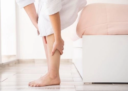
Inferior Vena Cava Syndrome occurs when there is a blockage or compression of the large vein (inferior vena cava) that collects blood from the lower part of the body and returns it to the heart. This blockage affects blood flow and causes symptoms like leg swelling and pain.
Main causes of IVCS:
Blood clots (Inferior Vena Cava thrombosis):
This is the most common cause, and it can result from:
Conditions that increase blood clotting tendency.
Prolonged immobility, such as long-distance travel or extended bed rest.
Major surgeries affecting circulation.
Tumors pressing on the vein:
Some cancers can compress the vein and cause obstruction, including:
Kidney cancer (especially if it invades the vein).
Liver tumors.
Uterine or ovarian tumors.
Tumors located behind the peritoneum (retroperitoneal area).
Pregnancy:
Especially in late pregnancy, the enlarged uterus compresses the inferior vena cava, impeding blood flow.
Heart or liver failure:
If the heart or liver function is impaired, blood can accumulate in the veins of the legs, reducing normal drainage.
Congenital causes:
Some people are born with a malformation or narrowing of the inferior vena cava that may only manifest later or under certain conditions.
Infections or direct injury:
Such as:
Infections caused by venous catheterization.
Injuries or surgical trauma in the abdomen or pelvis.
The inferior vena cava collects blood from the lower body — legs, pelvis, and abdomen — and returns it to the heart. When this vein is blocked or compressed, blood pools in the lower body, causing noticeable and uncomfortable symptoms.
Common symptoms:
Leg swelling:
Usually starts from the feet and progresses upwards, possibly affecting both sides or reaching the thighs.
Leg pain or heaviness:
A feeling of discomfort or pressure in the legs that worsens with prolonged standing or walking.
Visible varicose veins:
Enlarged veins visible under the skin, especially around the abdomen near the navel or in the groin.
Skin discoloration:
The skin may appear red or bluish due to poor blood flow.
Deep vein thrombosis (DVT):
Blood clots in the legs may cause or result from the blockage, requiring urgent treatment.
Swelling of the genital area:
If the obstruction extends to the pelvis, swelling of the genital organs in men or women may occur.
When IVCS is suspected, the doctor follows systematic steps to identify the cause, location of blockage, and if complications are present:
Clinical examination:
History of symptoms like leg swelling, pain, varicose veins, or skin changes.
Past medical history including previous clots, abdominal or pelvic tumors, or pregnancy.
Physical examination of the legs to check for swelling and prominent veins on the abdomen or groin.
Imaging and tests:
Doppler Ultrasound:
The first and simplest test to detect clots in deep or superficial veins.
CT scan with contrast:
Important to localize the obstruction or tumor pressing on the vein and to evaluate abdominal and pelvic masses.
Magnetic Resonance Imaging (MRI):
Used if there is suspicion of tumor or congenital abnormalities.
Venography (Contrast Venogram):
A detailed test where dye is injected into the vein to visualize blood flow and pinpoint the blockage. Not routinely done but very precise.
Blood tests:
Including D-dimer test to check for clot presence and other blood work to evaluate clotting tendencies.
If untreated or diagnosed late, IVCS may lead to serious complications:
Progressive deep vein thrombosis (DVT):
Clots can enlarge, block larger veins, worsening obstruction.
Pulmonary embolism:
If part of a clot breaks off and travels to the lungs, blocking pulmonary arteries—a life-threatening emergency.
Severe and chronic leg swelling:
Affecting mobility and quality of life, often associated with persistent pain.
Skin ulcers:
Poor blood flow causes wounds or ulcers that are difficult to heal.
Chronic venous congestion:
Long-term blood pooling leads to vein dilation and painful varicose veins.
Tissue fibrosis around the vein:
Over time, surrounding tissues may harden and scar, complicating treatment and worsening the condition.
Treatment depends on the underlying cause of the blockage or compression and the severity of symptoms.
Treating the underlying cause:
If caused by a blood clot (DVT):
Anticoagulant (blood-thinning) medications are prescribed to prevent new clots and help dissolve existing ones. Duration varies, sometimes lasting several months.
If caused by a tumor or external compression:
Treatment of the tumor with chemotherapy, radiation, or surgery, depending on tumor type and size.
If due to pregnancy or other factors:
Treatment targets the specific cause, such as careful monitoring during pregnancy or managing other health issues.
Supportive treatment to relieve symptoms:
Leg elevation:
Sitting or lying with legs raised helps reduce swelling and improves blood return to the heart.
Compression stockings:
Special stockings apply gentle pressure on the legs to reduce swelling and prevent new clots.
Painkillers and anti-inflammatory drugs:
Used to relieve pain and inflammation if needed.
Advanced medical procedures:
Angioplasty and stenting:
In some cases, stents are inserted into the vein to widen the blocked area and improve blood flow.
Thrombectomy (removal of clots) may be performed if clots are large.
Surgery:
Rarely used, reserved for severe blockages or cases not responding to medications and less invasive treatments.
4. Prevention and Follow-Up
Regular follow-up with the doctor to monitor the condition and adjust treatment if needed.
Lifestyle modifications: maintaining a healthy weight, exercising regularly, and avoiding prolonged sitting or standing.
In some cases, the patient may need to take anticoagulant medications for a long period to prevent recurrent clots.
Types of Surgeries for Treating Vena Cava Problems (Superior or Inferior)
Issues of blockage or compression in the vena cava may sometimes require surgical intervention. The choice of surgery depends on the patient’s condition and severity of the problem.
Thrombectomy (Clot Removal)
This surgery removes the blood clot blocking the vena cava.
It can be done via open surgery or minimally invasive techniques.
It’s mostly used in emergencies or when the clot is large and causes severe symptoms.
Venous Angioplasty and Stenting (Opening and Widening the Blockage)
The doctor inserts a medical balloon to widen the narrowed or blocked part of the vein.
Then, a metal stent is placed to keep the vein open and allow blood flow.
This is a less invasive method and has become common especially for chronic blockages.
Venous Bypass Surgery
When widening or stenting is not possible, a bypass is created by connecting another blood vessel (from a natural vein or synthetic graft) to bypass the blocked segment.
This is a major surgery used in complex cases.
Venous Resection and Reconstruction
In cases where the vein is severely damaged due to tumors or injuries, the damaged segment is removed.
Then, reconstruction is done using another blood vessel or synthetic graft.
This is a delicate surgery requiring high expertise.
Decompression Surgery
If the vein is compressed by tumors, enlarged lymph nodes, or inflammation, surgery may be needed to relieve the pressure.
This may include removing the tumors or tissues pressing on the vein.
Superior Vena Cava (SVC) Reconstruction Surgery
Specifically for treating problems of the superior vena cava caused by compression or blockage due to chest tumors or other reasons.
It involves removing the affected segment and reconstructing the vein to ensure normal blood return to the heart.
Endovascular Stenting (Catheter and Internal Stent Placement)
This is not an open surgery but an important interventional procedure.
Thin tubes (catheters) are inserted through blood vessels to place stents inside the vein to widen the blocked or narrowed area.
The advantage is less pain and faster recovery compared to traditional surgery.
Placement of Filters inside the Inferior Vena Cava (IVC Filters)
A filter (like a cava filter) is placed inside the inferior vena cava to prevent blood clots from reaching the lungs.
This is not a treatment for the blockage itself but an important preventive procedure, especially in patients at high risk for pulmonary embolism.


